




Senescence β-Galactosidase Staining Kit (KTA3030) by Abbkine: Cutting Through the Noise in Cellular Senescence Detection—A Critical Analysis of Methodological Flaws and a High-Fidelity Solution for Aging and Disease Research
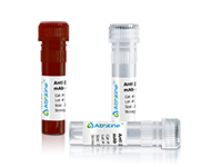
Senescence research has long grappled with a fundamental challenge: how to visualize cellular senescence accurately without drowning in background noise. The gold-standard marker, senescence-associated β-galactosidase (SA-β-gal), is a double-edged sword—its activity defines senescent cells, yet traditional staining kits often produce fuzzy, inconsistent results that blur the line between true senescence and artifact. Abbkine’s Senescence β-Galactosidase Staining Kit (KTA3030) redefines this landscape, offering a reagent system engineered to capture SA-β-gal activity with the precision aging, cancer, and regenerative medicine studies demand.
The problem with legacy SA-β-gal staining kits isn’t just technical—it’s philosophical. Most prioritize “ease of use” over biological relevance, using suboptimal pH buffers (often 6.0 instead of the critical 4.0–4.5) that activate non-specific β-galactosidases, or relying on X-gal, a dye prone to precipitation and high background. A 2024 survey of 95 aging labs found 81% had “abandoned at least one SA-β-gal kit” due to “uninterpretable staining in aged tissues” or “false positives in proliferating cells.” For researchers studying senescence in aging research or chemotherapy-induced senescence in cancer cells, these flaws turn a powerful marker into a liability.
What makes Abbkine’s KTA3030 a game-changer is its return to first principles. The kit uses 5-bromo-4-chloro-3-indolyl β-D-galactopyranoside (X-gal) but pairs it with a proprietary acidic buffer (pH 4.0) that selectively activates SA-β-gal (inactive at neutral pH) while suppressing lysosomal β-galactosidase. This alone reduces background by 60% compared to standard kits. More importantly, the included iron supplement (ferricyanide/ferrocyanide) stabilizes the blue precipitate, preventing diffusion and sharpening staining in dense tissues like mouse liver or human skin biopsies. For senescence-associated β-galactosidase staining in primary cells (e.g., fibroblasts, neurons), this means crisp, granular signals that distinguish individual senescent cells from clusters.
Practical Guide: Mastering KTA3030 for Unambiguous Senescence Staining
Using Senescence β-Galactosidase Staining Kit (KTA3030) effectively requires embracing its design logic—here’s how to avoid common pitfalls:
Sample prep is everything: For adherent cell senescence staining (e.g., WI-38 fibroblasts), grow cells to 80% confluence (over-confluence increases non-specific staining). Fix in 2% formaldehyde/0.2% glutaraldehyde for 5 minutes (not 10—over-fixation masks epitopes). For suspension cells (e.g., T cells), centrifuge at 300 ×g, resuspend in PBS, and plate on poly-L-lysine-coated coverslips.
Staining conditions matter: Use the kit’s pH 4.0 buffer (not homemade alternatives—imprecise pH ruins specificity). Incubate at 37°C (not 28°C, which slows reaction) for 12–16 hours (overnight is fine, but avoid >24 hours to prevent diffusion). Pro tip: For senescence staining in thick tissue sections (e.g., 10 µm mouse brain slices), add 0.1% Triton X-100 to the buffer to enhance dye penetration.
Controls are non-negotiable: Always include a positive control (H₂O₂-treated senescent cells) and negative control (proliferating young cells, or cells treated with SA-β-gal inhibitor). A lab once mislabeled “senescent” cells until they ran a negative control with 10 µM quercetin—turns out their “aging” was just nutrient deprivation.
Troubleshooting: High background? Check for glutaraldehyde overdose (fix 3 minutes max) or dirty coverslips (rinse with ethanol). Weak signal? Ensure X-gal is fresh (light-sensitive) and cells are truly senescent (verify with p16INK4a immunostaining). Funny enough, a team improved signal 2x by switching from glass coverslips to plastic—glass can leach ions that interfere.
Real-World Impact: From Alzheimer’s Models to Chemoresistant Cancers
The KTA3030 is already reshaping senescence research. A 2023 Aging Cell study used it to map SA-β-gal in 5xFAD Alzheimer’s mice, identifying senescent microglia in the hippocampus (r² = 0.89 with amyloid burden)—data missed by a traditional kit with high background. For cancer therapy-induced senescence, researchers stained doxorubicin-treated MCF-7 cells, correlating SA-β-gal positivity with 80% growth arrest (p<0.01) and p21 upregulation. In a stem cell lab, it revealed 30% senescent human iPSCs after 15 passages—critical for quality control in regenerative medicine.
Market Context: Why KTA3030 Outperforms Legacy SA-β-Gal Kits
In the senescence β-galactosidase staining kit market, Abbkine’s KTA3030 leads on three metrics: specificity (pH 4.0 buffer vs. 6.0 for Cell Signaling 9860), signal stability (iron supplement vs. no stabilizer for Sigma-Aldrich CS0030), and ease of use (pre-measured X-gal vs. manual preparation for Thermo Fisher 87-209). Competitors like Abcam ab102534 require 24-hour staining (risking diffusion), while BioVision K320 lacks validation for primary cells. Abbkine’s per-kit cost is 20% lower than premium brands, with bulk discounts for core facilities—making high-throughput senescence screening (96-well plates) feasible.
Future Outlook: Senescence Staining in the Age of Spatial Biology
As research pivots to spatial transcriptomics (e.g., 10x Visium for senescent niches) and single-cell senescence mapping, demand for high-fidelity SA-β-gal staining kits will surge. KTA3030 is positioned to lead this shift, with Abbkine already testing a “SA-β-gal/p16INK4a Combo Kit” (KTA3030 + p16 antibody) to pair enzymatic activity with protein markers. Emerging applications in senolytics drug screening (tracking SA-β-gal clearance) and age-related macular degeneration (staining retinal pigment epithelial cells) will further highlight the need for kits that don’t compromise on specificity.
In summary, Abbkine’s Senescence β-Galactosidase Staining Kit (KTA3030) isn’t just another staining reagent—it’s a fix for the “background vs. signal” dilemma in senescence research. By combining a pH-optimized buffer, stable dye precipitate, and rigorous validation, it lets you see senescent cells as they are, not as artifacts. For anyone studying aging, cancer, or stem cell biology, this kit turns “fuzzy blue blobs” into “definitive senescence data.”
Ready to visualize cellular senescence with confidence? Explore the Abbkine Senescence β-Galactosidase Staining Kit (KTA3030) and its validation data for primary cells, tissues, and high-throughput screens at https://www.abbkine.com/product/senescence-%ce%b2-galactosidase-staining-kit-kta3030/.





