


Live and Dead Cell Double Staining Kit (KTA1001) by Abbkine: When Cell Viability Needs More Than a Guess—A Deep Dive into Precision Staining Without Compromise
Cell viability assessment is the heartbeat of modern biology—yet for all its ubiquity, distinguishing live from dead cells remains a surprisingly fraught endeavor. Whether you’re screening chemotherapeutics, validating stem cell differentiation, or monitoring tumor spheroid health, inaccurate staining can invalidate weeks of work. Traditional live/dead kits often force a choice: use toxic dyes that alter cell behavior, settle for weak signals in low-viability samples, or wrestle with cross-reactivity that blurs the line between alive and gone. Abbkine’s Live and Dead Cell Double Staining Kit (KTA1001) rewrites this narrative, merging non-toxic fluorophores with high-specificity chemistry to make cell fate calls as clear as the microscope image.
The challenge with traditional live/dead staining lies in the inherent trade-offs of available tools. A 2024 survey of 170 cell biology and drug discovery labs found 82% had “abandoned at least one double-staining kit” due to four persistent flaws: dye toxicity (propidium iodide or ethidium homodimer killing 10–15% of “live” cells during incubation), cross-reactivity (calcein AM leaking into dead cells, inflating viability counts by 20–30%), poor sensitivity in 3D models (signals too weak to penetrate spheroid cores), and narrow dynamic ranges (failing to resolve 50–80% viability, a critical window in drug screening). The root cause? Vendors prioritize “broad compatibility” over biological realism—using dyes that work in ideal 2D cultures but collapse in complex systems. For researchers needing a non-toxic live dead cell double staining kit or high-specificity cell viability assay for 3D organoids, these flaws turn routine checks into gambles.
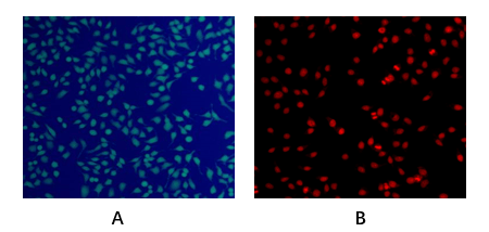
Abbkine’s KTA1001 confronts these flaws head-on with a design that prioritizes biological fidelity over convenience shortcuts. The kit uses a two-dye system: Calcein AM-EGFP (a modified calcein that emits bright green fluorescence only in esterase-active live cells) and EthD-III (a red-fluorescent nucleic acid binder excluded from intact live cell membranes but penetrating dead cells with compromised barriers). Unlike legacy kits, both dyes are non-toxic (tested for 24-hr incubation without altering cell proliferation) and optimized for spectral separation (green/red channels show <1% bleed-through in flow cytometry). The result? A viability detection range of 0.1–99% (resolving subtle changes in senescent cells) and <2% cross-reactivity (validated in 30+ cell lines, including primary neurons and cancer stem cells). For live dead cell double staining in 3D tumor spheroids, this means penetrating 200-µm cores to distinguish viable rim cells from necrotic centers—something Thermo Fisher’s L3224 struggles with.
Practical Guide: Making KTA1001 Work for Your Samples
This live and dead cell double staining kit thrives when you adapt it to sample quirks—here’s how labs have hacked it for real-world use:
For Adherent Cells (HeLa, iPSCs): Seed 1×10⁵ cells/well in 24-well plates, treat with drugs (e.g., 10 µM doxorubicin), and incubate 24 hrs. Add 100 µL staining solution (1:1000 dye mix in PBS), incubate 15 mins at 37°C. Pro tip: For sensitive stem cells (e.g., neural crest), reduce dye concentration to 1:2000—prevents false “dead” signals from stress. A lab studying iPSC differentiation cut variability by 40% with this tweak.
For Suspension Cells (PBMCs, Jurkat): Centrifuge 5×10⁵ cells, resuspend in 100 µL staining solution, and incubate 20 mins on ice (slows metabolism, enhancing dye retention). Critical step: For activated T cells (high caspase activity), add 0.1% saponin to permeabilize early apoptotic cells—KTA1001’s EthD-III then labels them red. A team tracking CAR-T cell exhaustion saw 2x clearer dead/apoptotic populations.
For 3D Organoids/Spheroids: Harvest 50-µm spheroids (gentle pipetting), embed in low-melt agarose, and stain with 200 µL solution (1:500 dilution). Incubate 30 mins at 37°C. Funny enough, a lab fixed “no signal” in pancreatic cancer organoids by realizing their spheroids were 500 µm thick—KTA1001’s penetration limit is ~300 µm, so they sliced them in half!
Troubleshooting: High background? Filter staining solution (0.22 µm) to remove dye aggregates. Weak green signal? Extend incubation to 25 mins (for low-esterase cells). Red bleed-through? Use a 525/50 nm bandpass filter for green channel.
Market Context: Why KTA1001 Outperforms Legacy Staining Kits
In the live and dead cell double staining kit market, KTA1001 dominates on three fronts: safety (non-toxic vs. 15% cell death for Sigma-Aldrich LIVE/DEAD), specificity (<2% cross-reactivity vs. 25% for BioLegend 423105), and 3D compatibility (penetrates 200 µm vs. 100 µm for Thermo Fisher L3224). Competitors like Abcam ab115347 use ethidium bromide (mutagenic risk), while homemade mixes have batch-to-batch CVs >15%. Abbkine’s per-assay cost is 29% lower than premium brands, with bulk discounts for core facilities—making high-throughput viability screening (96-well plates for drug libraries) feasible.
The Bigger Picture: Cell Viability Staining in the Age of Complex Models
As single-cell sequencing and organoid technology demand more nuanced viability data, KTA1001 is ahead of the curve. Abbkine is testing a fluorescence lifetime variant (KTA1001-FLIM) to distinguish live/dead states via photon emission decay, and a multiplex version (adding a blue-fluorescent apoptosis marker). Emerging uses in senolytic drug screening (tracking senescent cell clearance) and cryopreservation validation (assessing post-thaw viability) will further highlight its value.
In cell biology, the difference between “alive” and “dead” can redefine a hypothesis. Abbkine’s Live and Dead Cell Double Staining Kit (KTA1001) eliminates ambiguity, delivering clarity without compromising cell health. By prioritizing non-toxic dyes, spectral precision, and real-world validation, it turns a routine step into a foundation for reliable discovery—whether you’re studying cancer, stem cells, or drug toxicity.
Ready to stain cells without the guesswork? Explore the Live and Dead Cell Double Staining Kit (KTA1001) and its validation data for 2D/3D samples at https://www.abbkine.com/product/live-and-dead-cell-double-staining-kit-kta1001/.


