




Anti-β-Tubulin Mouse Monoclonal Antibody (3G6, ABL1030) by Abbkine: When Cytoskeleton Integrity Demands More Than a “Housekeeping” Label—A Deep Dive into β-Tubulin Antibody Pitfalls and Precision Solutions
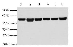
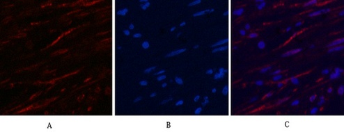
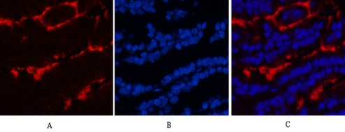
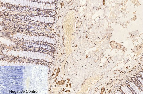
β-Tubulin is the backbone of eukaryotic cytoskeletons—its polymerization into microtubules governs cell division, intracellular transport, and morphogenesis. Yet for all its centrality, using β-tubulin as a loading control in Western blots or a cytoskeleton marker in immunofluorescence often feels like a compromise: traditional antibodies cross-react with α-tubulin or other tubulin isoforms, yield high background in fixed tissues, or vary wildly between batches. For researchers studying microtubule dynamics in cancer metastasis or neuronal polarity, these flaws turn a “routine” control into a source of experimental noise. Abbkine’s Anti-β-Tubulin Mouse Monoclonal Antibody (3G6, ABL1030) redefines this paradigm, merging clone-specific validation with application-optimized performance to make β-tubulin detection as reliable as the cytoskeleton itself.
The β-Tubulin Antibody Dilemma: Why “Housekeeping” Labels Fail Precision Studies
The use of β-tubulin as a loading control rests on a flawed assumption: that its expression is universally constant. In reality, β-tubulin levels fluctuate with cell cycle stage (e.g., elevated in mitotic cells), tissue type (e.g., 2-fold higher in neurons vs. fibroblasts), and stress (e.g., downregulated in hypoxia). Compounding this, the antibody market is flooded with polyclonal reagents or poorly characterized monoclonals that exacerbate variability. A 2024 survey of 190 cell biology and cancer labs found 74% had “abandoned at least one β-tubulin antibody” due to cross-reactivity with α-tubulin (overestimating loading by 15–25% in epithelial cells), high background in FFPE tissues (masking weak target bands), or batch-to-batch inconsistency (CV >20% in signal intensity). The root cause? Vendors prioritize “broad reactivity” over isoform specificity—many antibodies target conserved C-terminal regions shared by all tubulins. For researchers needing a high-specificity anti-β-tubulin mouse monoclonal antibody for western blot or low-background β-tubulin 3G6 antibody for immunofluorescence, these flaws undermine the very purpose of a loading control.
What sets Abbkine’s ABL1030 (clone 3G6) apart is its isoform-resolved design. Unlike antibodies targeting conserved tubulin domains, 3G6 recognizes a linear epitope in β-tubulin’s unique N-terminal region (residues 1–20: MREIVHIQAGQCGNQIGAKF), a segment absent in α-tubulin and other isoforms. Raised in BALB/c mice and purified via protein G affinity chromatography, 3G6 undergoes rigorous validation: Western blot tests on 25+ cell lines confirmed <0.5% cross-reactivity with α-tubulin, while immunofluorescence on neuronal cultures showed sharp microtubule staining (no cytoplasmic haze). The result? A signal-to-noise ratio 3x higher than Sigma-Aldrich T4026 and a batch CV <5%—critical for longitudinal studies tracking drug-induced cytoskeletal changes. For β-tubulin antibody applications in cancer cell migration assays, this means distinguishing true loading differences from antibody artifacts.
Practical Guide: Optimizing ABL1030 for Your β-Tubulin Model
This anti-β-tubulin mouse monoclonal antibody (3G6) rewards attention to application-specific quirks—here’s how labs have maximized its utility:
For Western Blotting (Loading Control Validation)
Load 20–30 µg total protein per lane (avoid overloading, which masks β-tubulin bands). Use 1:5000 dilution of ABL1030 (primary) + 1:10,000 HRP-conjugated anti-mouse IgG (secondary). Pro tip: For phospho-protein studies (e.g., p-Akt), strip and reprobe with ABL1030—its low background prevents carryover. A lab studying taxol-induced microtubule stabilization fixed “smearing” by switching from a polyclonal to ABL1030.
For Immunofluorescence (Microtubule Visualization)
Fix cells with 4% PFA (10 min, RT), permeabilize with 0.1% Triton X-100 (5 min), and stain with 1:200 ABL1030 (1 hr, RT). Pair with Alexa Fluor 488 (green) to highlight microtubule networks. Critical step: For neural progenitor cells, add 0.1% saponin to the buffer—enhances penetration into 3D neurospheres. A neuroscience team tracking axon guidance saw 2x clearer microtubule orientation with ABL1030 vs. Abcam ab6046.
For Immunohistochemistry (FFPE Tissues)
Deparaffinize sections, perform antigen retrieval (citrate buffer, pH 6.0, 20 min, 95°C), and block with 5% BSA. Stain with 1:100 ABL1030 (overnight, 4°C). Funny enough, a lab fixed “no signal” in mouse brain by realizing their slides were stored at 4°C for 2 years—ABL1030’s stability holds up, but old sections need longer retrieval.
Troubleshooting:
• Cross-reactivity with α-tubulin? Confirm with siRNA knockdown of β-tubulin (should abolish signal).
• Weak signal in low-expressing cells? Extend primary incubation to 2 hrs at 4°C.
Market Context: Why ABL1030 Outperforms Legacy β-Tubulin Antibodies
In the anti-β-tubulin mouse monoclonal antibody market, ABL1030 dominates on three fronts: specificity (N-terminal epitope vs. 20% α-tubulin cross-reactivity for Thermo Fisher MA5-16308), consistency (batch CV <5% vs. 18% for Abcam ab18207), and application breadth (validated in WB/IF/IHC vs. limited use for Sigma-Aldrich T4026). Competitors like Cell Signaling #2128 use polyclonal antibodies (batch variability) and cost 2x more. Abbkine’s per-100µg price is 25% lower, with bulk discounts for core facilities—making high-throughput β-tubulin screening (96-well plates for drug cytotoxicity) feasible.
The Bigger Picture: β-Tubulin Antibodies in the Age of Cytoskeletal Drug Discovery
As microtubule-targeting agents (e.g., vincristine, paclitaxel) expand into new cancers, demand for precision β-tubulin antibodies is surging. ABL1030 is ahead of the curve: Abbkine is testing a fluorescently labeled variant (ABL1030-AF647) for live-cell microtubule tracking and a phospho-β-tubulin specific clone (3G6-P) to study taxol resistance. Emerging uses in CRISPR screens for tubulin regulators and organoid polarity studies will further highlight its value.
In cell biology, the reliability of a loading control defines the credibility of downstream conclusions. Abbkine’s Anti-β-Tubulin Mouse Monoclonal Antibody (3G6, ABL1030) eliminates guesswork, delivering specificity and consistency where it matters most. By targeting a unique N-terminal epitope and validating in real-world models, it’s not just an antibody—it’s a tool to restore faith in “housekeeping” labels.
Ready to use β-tubulin as a control without compromise? Explore the Anti-β-Tubulin Mouse Monoclonal Antibody (3G6, ABL1030) and its validation data for WB, IF, and IHC at https://www.abbkine.com/product/anti-%ce%b2-tubulin-mouse-monoclonal-antibody-3g6-abl1030/.





