| Product name | Anti-β-Tubulin Mouse Monoclonal Antibody (3G6) |
| Immunogen | Synthetic Peptide |
| Host | Mouse |
| Reactivity | Chicken, Dog, Hamster, Human, Insect, Monkey, Mouse, Rabbit, Rat, Sheep, Yeast |
| Applications | IF, IHC-P, WB |
| Applications notes | Optimal working dilutions should be determined experimentally by the investigator. Suggested starting dilutions are as follows: WB (1:10000), IHC-P (1:400), IF (1:400). |
| Clonality | Monoclonal |
| Preparation method | The antibody was affinity-purified from mouse ascites by affinity-chromatography using specific immunogen |
| Alternative | TUBB3; TUBB4; Tubulin beta-3 chain; Tubulin beta-4 chain; Tubulin beta-III |
| Formulation | Liquid solution |
| Concentration | 1 mg/ml |
| Storage buffer | Liquid in PBS, pH 7.4, containing 0.02% Sodium Azide as preservative and 50% Glycerol. |
| Storage instructions | Stable for one year at -20°C from date of shipment. For maximum recovery of product, centrifuge the original vial after thawing and prior to removing the cap. Aliquot to avoid repeated freezing and thawing. |
| Shipping | Gel pack with blue ice. |
| Precautions | The product listed herein is for research use only and is not intended for use in human or clinical diagnosis. Suggested applications of our products are not recommendations to use our products in violation of any patent or as a license. We cannot be responsible for patent infringements or other violations that may occur with the use of this product. |
| Background | Tubulin is one of several members of a small family of globular proteins. The most common members of the tubulin family are α-tubulin and β-tubulin, the proteins that make up microtubules. Each has a molecular weight of approximately 55 kDa. Antibodies against β-Tubulin are useful as loading controls for Western Blotting. However it should be noted that levels of β-Tubulin may not be stable in certain cells, such as adipose tissue. |
| Gene ID | 10381 |
| Alternative | TUBB3; TUBB4; Tubulin beta-3 chain; Tubulin beta-4 chain; Tubulin beta-III |
| Accession | Q13509 |
| Observed Band(KD) | 55 |
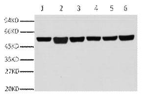
Fig.1. Western blot analysis of A549(1), rat brain (2), mouse brain (3), chicken lung (4) and rabbit testis(5), sheep muscle(6), diluted at 1:10000.
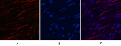
Fig.2. Immunofluorescence analysis of human appendix tissue. 1, β-Tubulin Monoclonal Antibody (3G6) (red) was diluted at 1:400 (4°C, overnight). Picture A: Target. Picture B: DAPI. Picture C: merge of A+B.
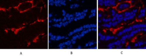
Fig.3. Immunofluorescence analysis of mouse lung tissue. 1, β-Tubulin Monoclonal Antibody (3G6) (red) was diluted at 1:400 (4°C, overnight). Picture A: Target. Picture B: DAPI. Picture C: merge of A+B.
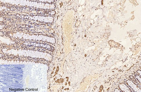
Fig.4. Immunohistochemical analysis of paraffin-embedded human colon tissue. 1, β-Tubulin Monoclonal Antibody (3G6) was diluted at 1:400 (4°C, overnight). Negative control was used by secondary antibody only.
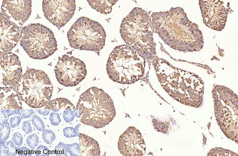
Fig.5. Immunohistochemical analysis of paraffin-embedded mouse testis tissue. 1, β-Tubulin Monoclonal Antibody (3G6) was diluted at 1:400 (4°C, overnight). Negative control was used by secondary antibody only.
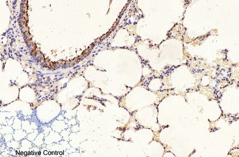
Fig.6. Immunohistochemical analysis of paraffin-embedded rat lung tissue. 1, β-Tubulin Monoclonal Antibody (3G6) was diluted at 1:400 (4°C, overnight). Negative control was used by secondary antibody only.
Author:Lv, Meinan, et al Publication name:Journal of the American Chemical Society 138 IF:15
Author:Zhang, Yu, et al Publication name:Journal of Experimental & Clinical Cancer Research IF:13
Author:FC Huo, ZM Zhu, WQ Du, YJ Pan, X Jiang Publication name:Pharmacological Research IF:9
Author:Cai B, Ma M, Yuan R Publication name:Cellular & Molecular Biology Letters IF:8
Author:**ong J, Wang L, Feng Z Publication name:Antioxidants IF:7
Author:Fang, Ting, et al. Publication name:Microbiological Research IF:6.9
Author:Lu, Hui, et al. Publication name:Mbio IF:6
Author:X Tan, X Wang, X Liao, X Wang, Z Jiang, W Liang Publication name:Iscience IF:6
Author:Xiong XX, Pan F, Chen RQ, et al Publication name:Cell Death Dis IF:6
Author:Lin, Yao, et al. Publication name:Cells IF:5.2
Author:Zou X, Chen C, Huang X, et al Publication name:Talanta IF:5
Author:SK Kim, GY Lee, SK Kim, YJ Kwon Publication name:Molecular Neurobiology IF:5
Author:Bi, Jian, Yufen Wang, and Yingde Wang. Publication name:Frontiers in Pharmacology IF:4.8
Author:Liu, Jialin, et al. Publication name:Biomedical and Environmental Sciences IF:4.1
Author:Sun, Shiquan, et al. Publication name:BMC cancer IF:4
Author:Chen, Jiaxi, et al. Publication name:Toxicology and Applied Pharmacology IF:4
Author:Ye, Jia-Xin, et al Publication name:American Journal of Physiology-Lung Cellular and Molecular Physiology 310 IF:4
Author:Li, Xiaojun, et al Publication name:Virus Research 227 (2017): 240-244 IF:3
Author:Zhang L, Chen P, Yang S, et al Publication name:Oncology Letters IF:2
Author:Zeng T, Liu F, Zhou J, et al Publication name:Endocrine journal IF:2
Author:Lu Y, Lin B, Zhong J, et al Publication name:Biological & Pharmaceutical Bulletin IF:2
Author:Li J, Ma Y, Yuan W, Xiao Y, et al Publication name:Insect Biochem Mol Biol IF:1
You must be logged in to post a review.


Reviews
There are no reviews yet.