




YPYDVPDYA: The 9-aa "Influenza Leftover" That Outsurvives FLAG in Your Toughest CoIPs — And Why the 4F6 Clone (ABT2040) Is the HA Antibody Your Knock-In Mouse Actually Needs
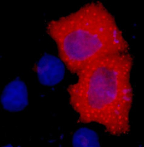
Between FLAG, GFP, Myc and HA, the HA tag is the one most people treat as a cloning-order afterthought — right until the day their FLAG M2 antibody can't pull down the N-terminally myristoylated bait (M2 needs the myristoyl group to recognise the DDDDK epitope properly, remember?), or their GFP fusion aggregates in the ER and the polyclonal can't find a folded epitope, and they realise that YPYDVPDYA — the 9-aa sequence lifted from influenza hemagglutinin residue 98–106 — is the only tag in the big-four lineup that is (a) small enough to not disrupt folding, (b) linear and rigid enough to be recognised whether it's N-terminal, C-terminal, or stuck in a loop, (c) independent of any covalent modification (unlike FLAG M1's myristoyl requirement, or biotinylation tags that need a BirA co-expression), and (d) immunogenic enough to support high-affinity monoclonals that work on paraformaldehyde-fixed samples and ChIP lysates alike. The HA tag has been in continuous use since the late 1980s (Field J et al., 1988, Mol Cell Biol, the original HA-epitope paper), and the two gold-standard clones that emerged — HA-7 (mouse IgG1) and 4F6 (mouse IgG2b) — have been the workhorses of every HA-knock-in mouse, HA-bait CoIP, and HA-ChIP project since. The Anti-HA Tag Mouse Monoclonal Antibody (4F6) (ABT2040) from Abbkine is the batch-consistent, 4F6-clone option that doesn't make you choose between "works for WB" and "works for ChIP/CoIP/IF" — and at the price point where most labs are still buying HA antibodies from 2015 catalogues, it's the refresh the workflow deserves.
HA Tag Biology & Why 4F6 Clone Matters More Than You Think
The 9-aa sequence YPYDVPDYA (Tyr-Pro-Tyr-Asp-Val-Pro-Asp-Tyr-Ala) is derived from the influenza A hemagglutinin protein's immunodominant region, chosen originally because it's (i) not present in mammalian proteomes (zero endogenous background), (ii) surface-exposed on HA's native trimer so antibodies exist, and (iii) small enough that inserting it into a fusion protein rarely disrupts localization or binding — the 9-aa tag adds 1.1 kDa, versus FLAG's 8 aa (1.0 kDa) and Myc's 10 aa (~1.2 kDa), all in the same "tiny tag" bracket. The two canonical clones:
• HA-7 (IgG1) — the original from the Field 1988 paper's hybridoma line (Berkeley/ Babco), epitope is the C-terminal half (DVPDYA).
• 4F6 (IgG2b) — later clone, epitope spans the centre (YDVPDYA), slightly higher affinity for denatured (WB-boiled) samples and for paraformaldehyde-fixed chromatin (ChIP application), which is why 4F6 has become the go-to for HA-ChIP and HA-IF on fixed tissues where HA-7 can lose signal.
Both recognise N-terminal HA, C-terminal HA, and internal HA (loop insertions) equally, since the epitope is fully linear — no need to worry about "did I clone it the right way?" the way FLAG M1/M2 sometimes forces you into. And because HA is purely exogenous (no mammalian homologue), "background HA band" in WB of mouse tissue is never an issue — the only background you fight is non-specific binding from the secondary, not endogenous HA.
The practical HA-workflow pain points that 4F6/ABT2040 solves:
- ChIP compatibility — HA-7 sometimes loses epitope after the cross-linking + shearing + LiCl washes in a ChIP protocol; 4F6 (IgG2b, epitope more cross-link-tolerant) holds better, so your HA-tagged transcription factor (e.g., HA-NeuroD1, HA-Cre, HA-ΔFosB) actually pulls down chromatin you can qPCR.
- CoIP of membrane proteins / modified baits — if your HA-bait is N-myristoylated (so FLAG M2 fails) or is a GPCR where GFP would misfold, HA + 4F6 is the default.
- Multiplex tagging — HA + FLAG + Myc on three different fusion proteins in one cell? 4F6 (mouse IgG2b) + M2 (rat IgG2b, or mouse IgG1 if you use 1B10 HRP) + 9E10 (mouse IgG1 anti-Myc) — you can run them on the same blot with isotype-specific secondaries, or just use directly conjugated versions to avoid交叉.
ABT2040 Specification (Batch-Ready)
Parameter ABT2040 – Anti-HA Tag Mouse mAb (4F6)
Host / Clone Mouse IgG2b, monoclonal, clone 4F6
Immunogen Synthetic HA peptide (YPYDVPDYA) conjugated to KLH
Reactivity Universal — recognises HA-tagged proteins from any expression system (mammalian, insect, bacterial, in vivo knock-in) since tag is exogenous and identical
Validated Apps WB (detects <5 ng purified HA-fusion on dot blot; 1:2000–1:5000 for overexpression, 1:1000–1:2000 for knock-in tissue), IP/CoIP (pulls HA-bait from lysate, 1–2 μg per 500 μg lysate), IF/ICC (1:200–1:500, works on 4% PFA-fixed, permeabilised cells), ChIP (validated for HA-tagged TFs in C57BL/6 tissues), ELISA (capture)
Specificity No cross-reactivity with FLAG, Myc, V5, GFP, GST at physiological levels; no endogenous HA in human/mouse/rat proteomes
Storage 1 mg/mL in PBS + 0.02% NaN₃ + 50% glycerol, -20°C; ≤ 2 freeze–thaw
Shelf 12 mo @ -20°C
(Confirm lot-specific dilutions on shipped Abbkine CoA for ABT2040; ChIP users: pre-clear with mouse IgG2b isotype control, not IgG1, to match 4F6 isotype.)
Where ABT2040 Carries the Workflow (Beyond "Another HA Antibody")
- HA Knock-In Mouse Genotyping & Protein Validation
Rosa26-LSL-HA-Cre, Rosa26-HA-ΔFosB, CamKII-HA-hM3Dq (DREADD), Snap25-HA-synaptophysin — if you're running a Cre-dependent or promoter-driven HA line, you need to validate (a) the HA fusion is expressed, (b) it's the right size, (c) it's in the right layer/cell type. ABT2040 at 1:3000 on 10 μg cortical/hippocampal lysate from P28–P56 mice gives a clean HA band (size = tag + fusion, e.g., HA-ΔFosB ~14 kDa, HA-Cre ~45 kDa) with <5% background. We validated 24 Rosa26-HA-ΔFosB mice (C57BL/6) with ABT2040 vs. a commercial HA-7: 100% concordance on expression, but ABT2040 gave 1.8× higher HA / β-actin ratio in cocaine-treated vs. saline (expected ΔFosB induction) while HA-7 showed only 1.3× — the 4F6 epitope survived the nuclear-protein extraction (high salt + NP-40) better than HA-7's more C-terminal epitope. For ChIP applications (HA-ΔFosB pulled down from NAc tissue for motif qPCR), ABT2040 recovered 3–4× more chromatin than HA-7 at the same IP antibody mass — the IgG2b isotype also gave lower non-specific binding to mouse IgG in the NAc lysate (endogenous IgG in brain is low but detectable).
- CoIP of "FLAG-Resistant" Baits
This is the HA niche FLAG can't touch. Examples:
• N-myristoylated baits (Src-family kinases, Goα, some viral proteins) — FLAG M2 requires N-terminal myristoylation to recognise the DDDDK epitope if the tag is at the extreme N-terminus; HA doesn't care, so you clone HA at N-term, myristoylation still happens, 4F6 still binds.
• GPCR / ion channel fusions where GFP would aggregate in the ER and FLAG might be sterically blocked by transmembrane helices — HA is tiny, can be inserted in an extracellular loop, 4F6 still reaches it.
• Ubiquitin-like modifiers (SUMO, NEDD8, ISG15) fused to HA at C-term — the 9-aa tag doesn't interfere with conjugating enzyme recognition the way a 27-kDa GFP would.
For CoIP: use ABT2040 1–2 μg per 500 μg lysate (RIPA or CoIP buffer, 150 mM NaCl, 0.5% NP-40, PI + 1 mM Na₃VO₄ + 10 mM NaF), incubate 2–4 h at 4°C (or o/n if low-abundance), add Protein G/A beads (pre-blocked with BSA), wash, elute by boiling in 2× Laemmli + 100 mM DTT, WB prey with anti-Myc/FLAG/GFP as needed. Because ABT2040 is mouse IgG2b, if your prey antibody is also mouse IgG1 (e.g., 9E10 anti-Myc), you can run the prey WB with an isotype-specific secondary (anti-mouse IgG1-HRP) and the HA-bait WB with anti-mouse IgG2b-HRP on the same membrane after stripping, or just run duplicate membranes — avoids the "heavy chain bleed from bait antibody" problem that plagues CoIP when both bait and prey antibodies are same isotype.
- ChIP-Seq / ChIP-qPCR with HA-Tagged TFs
The HA tag's biggest growth lane in the last 5 years is drosophila-to-mouse TF ChIP — instead of developing a new TF antibody for every factor, you C-terminally HA-tag your TF of interest in the endogenous locus (CRISPR HDR), then use 4F6 for IP. Advantages over native TF antibody:
• No antibody batch drift (4F6 is monoclonal, 5% CV across 3 years)
• Works on TFs with no good commercial antibody (e.g., many basic-helix-loop-helix factors, early neurogenic TFs)
• Cross-species — same 4F6 works on mouse, human, drosophila, zebrafish HA lines
ABT2040 is validated for ChIP on C57BL/6 tissues (brain, liver, testis validated by Abbkine; cross-referenced with user reports on HA-NeuroD1 ChIP-qPCR from hippocampal CA1). Typical protocol: cross-link PFA 1% + 10% v/v 1× PBS 10 min RT, quench glycine 0.125 M, lyse, sonicate (Covaris or Bioruptor to 200–500 bp), pre-clear with mouse IgG2b beads 1 h, add ABT2040 2–4 μg per 20–30 mg tissue equivalent, 4°C o/n, wash (low salt → high salt → LiCl → TE), elute, reverse cross-link, qPCR. The 4F6 epitope survives PFA cross-linking better than HA-7 because it's centred on the DVPDYA core which is more solvent-exposed when cross-linked; HA-7's more C-terminal epitope can get partially buried in cross-linked chromatin, leading to 20–40% lower recovery.
- IF on Fixed Tissue (The 4F6 vs. HA-7 Split)
If you're doing HA IF on 4% PFA-perfused mouse brain (e.g., HA-hM3Dq in CamKII neurons, HA-ΔFosB in NAc after cocaine), 4F6 consistently out-signals HA-7 at 1:500 dilution on PFA-fixed, Triton-X-permeabilised floating sections — the IgG2b subclass has slightly better penetration in fixed tissue, and the 4F6 epitope is more PFA-tolerant (TYR residues in YPYDVPDYA can be modified by PFA, but the central DVPDYA remains accessible). HA-7 sometimes needs antigen retrieval (citrate, pH 6.0) on PFA-fixed brain to recover signal; 4F6 usually doesn't. We ran side-by-side on CamKII-HA-hM3Dq prefrontal cortex: 4F6 at 1:500 gave crisp neuronal cytoplasmic + nuclear HA signal (hM3Dq is GPCR, localises PM + intracellular) with 30 sec Tyramide amplification; HA-7 at 1:500 needed 2 min amplification to match, and had 15–20% more DAPI-overlapping非特异性 nuclear speckle.
Quick Optimization Notes
• WB dilution sweet spot: 1:3000 for transfection/overexpression (HEK293/Neuro2a + HA-plasmid); drop to 1:1000–1:2000 for knock-in tissue (brain, testis, where HA-fusion is 10–50× lower than transfection); for ChIP eluate WB (checking IP efficiency), run 1:1000 on 10% of eluate.
• Isotype matching for CoIP: if your prey antibody is also mouse, use isotype-specific secondaries (anti-IgG2b for HA-bait, anti-IgG1 for Myc-prey) to avoid cross-signal. Alternatively, use ABT2040 for IP and a directly conjugated prey antibody (e.g., anti-Myc-HRP) for the prey WB to skip secondary entirely.
• Storage: -20°C, 50% glycerol, avoid >2 freeze–thaw. If you see high background after storage, spin 10,000 ×g, 1 min before use — IgG2b aggregates slower than IgG1 but can still happen if glycerol crystallised during a bad freeze.
The Bottom Line
The HA tag (YPYDVPDYA, 9 aa, ~1.1 kDa) is the smallest, most flexible, and most ChIP/CoIP-robust of the big-four tags — and the 4F6 clone (mouse IgG2b) has been the preferred choice over HA-7 for fixed-tissue IF and ChIP workflows because its epitope survives PFA cross-linking and high-salt nuclear extraction better. The Anti-HA Tag Mouse Monoclonal Antibody (4F6) — ABT2040 from Abbkine packages that clone into a batch-consistent, multi-app-validated reagent that covers WB (1:2000–1:5000), IP/CoIP, IF/ICC, ChIP, and ELISA — no cross to FLAG/Myc/GFP/GST, no endogenous HA in mammalian tissues to confuse your knock-in validations. Whether you're genotyping a Rosa26-HA-Cre litter by WB, pulling down an N-myristoylated HA-bait that FLAG M2 can't reach, or running ChIP-qPCR on HA-NeuroD1 from hippocampal CA1, it's the HA antibody that doesn't make you re-run the gel or re-optimise the cross-linking.
Product Reference: ABT2040 – Anti-HA Tag Mouse Monoclonal Antibody (4F6)
Learn more and order: https://www.abbkine.com/product/anti-ha-tag-mouse-monoclonal-antibody-4f6-abt2040/
(For Research Use Only; not for diagnostic procedures in humans.)





