




Your TCA Cycle Paper Looks Great—Until the Reviewer Asks About FH Protein Levels. Here's Why Abbkine's ABM40073 Is the Antibody That Won't Embarrass You in Supplementary Figure 1
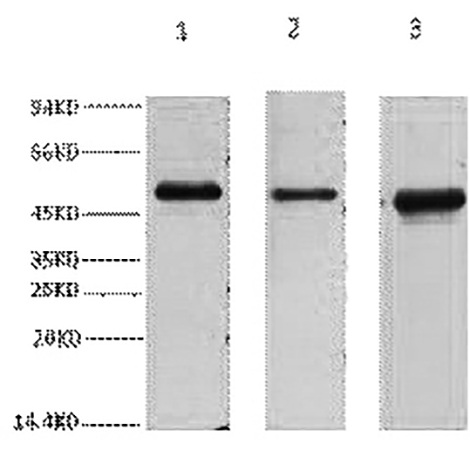
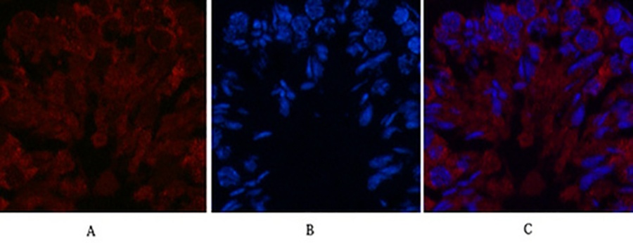
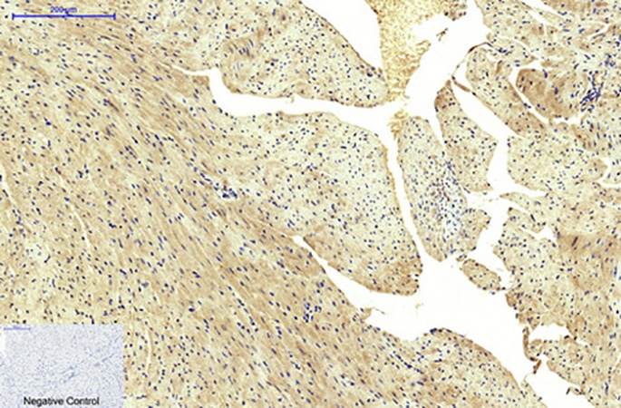
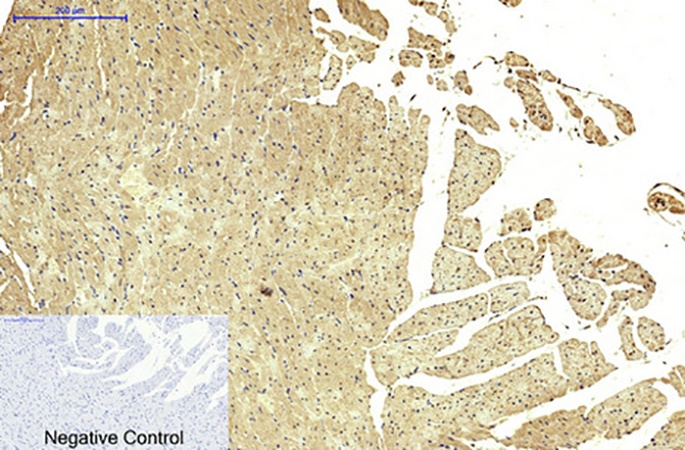
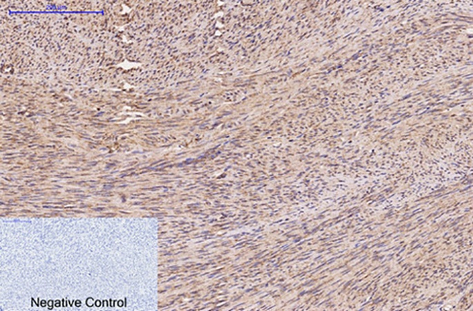
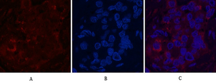
There is a very specific kind of panic among metabolism and cancer-epigenetics labs right now: you've built an elegant story around the Warburg effect, oncometabolite accumulation, or HLRCC-driven tumor suppression, your seahorse data and fumarate measurements look beautiful, and then the reviewer calmly asks you to actually show FH protein expression across your conditions with a "specific, validated antibody suitable for both Western blot and IHC." Suddenly your go-to β-actin loading control isn't the issue—your FH (Fumarate Hydratase / Fumarase) primary antibody is. Either it drags a smear across the 54 kDa region, lights up non-specifically in the cytosol, or simply refuses to work in paraffin sections, and your entire metabolic claim rests on a band that could be anything.
FH Isn't Just Another "Krebs Cycle Enzyme"—It's a Tumor Suppressor That Demands Respect
Fumarate Hydratase (FH, UniProt P07954, Gene ID 2271, EC 4.2.1.2) is the TCA-cycle enzyme that reversibly converts fumarate → L-malate—but calling it "just a metabolic housekeeper" is a profound undersell. In the last decade, FH has been reclassified as a bona fide tumor suppressor: germline FH mutations cause Hereditary Leiomyomatosis and Renal Cell Cancer (HLRCC), an aggressive type II papillary RCC syndrome, while somatic loss drives fumarate accumulation that acts as an oncometabolite—succinylation of proteins, stabilization of HIF-1α independent of oxygen, and epigenetic reprogramming via competitive inhibition of α-KG–dependent dioxygenases. In short: your FH readout isn't decorative. It's often the linchpin between "cells metabolically adapt" and "cells transform."
The catch? FH exists in two translated forms: a longer N-terminus-extended precursor targeted to mitochondria (where removal of the transit peptide yields the mature ~48–54 kDa homotetramer), and a cytosolic isoform initiated at a downstream start codon. Any antibody you use needs to recognize the conserved catalytic core shared across both compartments, be insensitive to minor denaturation differences between mitochondria-enriched vs. total lysates, and—critically—not cross-react with unrelated TCA enzymes of similar mass. That's where most generic "anti-FH/fumarase" polyclonals fail, and where a properly cloned monoclonal earns its catalog number.
Enter FH Monoclonal Antibody (ABM40073) by Abbkine — Precision Over Hope
Abbkine's FH Monoclonal Antibody (ABM40073) is a mouse IgG₁ monoclonal raised against a synthetic peptide / recombinant human FH protein immunogen, engineered for the exact applications where a polyclonal's background haze will sink you:
Parameter ABM40073 Specification
Target FH / Fumarate Hydratase (Fumarase) — UniProt P07954, Gene ID 2271
Antibody type Mouse Monoclonal (IgG₁) — monospecific epitope, superior batch consistency
Reactivity Human, Mouse, Rat (H, M, R cross-reactivity validated)
Applications WB (1:1,000–3,000) · IHC-P (1:50–200, paraffin) · IF/ICC (1:100–200)
Observed MW ~54 kDa (mature mitochondrial fumarase homotetramer subunit)
Format / Conc. Liquid, 1 mg/mL, in PBS pH 7.4 + 0.02% NaN₃ + 50% glycerol
Storage / Ship -20°C (1 year stable from ship date); aliquot to avoid freeze–thaw; ship blue-ice gel pack
Centrifuge before opening Yes — recommended to recover full contents before first use
The advantage of a monoclonal here isn't marketing fluff. It's three concrete things your reviewer cares about: (1) one defined epitope = one clean band at ~54 kDa (not a bouquet of non-specific ghosts); (2) lot-to-lot consistency that lets you reprobe a blot from samples extracted three months apart without "did the antibody change?" anxiety; and (3) the ability to go from a WB band in your lysate screen → straight into IHC-P on the same FFPE cohort with one antibody, one catalogue number, zero guesswork.
What Changes When Your FH Antibody Actually Behaves
① Your WB bands stop needing apologies.
With a recommended 1:3,000 WB dilution, you're not drowning the membrane in excess IgG that sticks everywhere. A clean ~54 kDa signal against 293T / HeLa / HepG2 / mouse tissue—with proper positive (FH-intact) and negative (FH-null HLRCC-line or siRNA-knockdown) controls—converts FH from "a band we think is right" to a defensible loading-normalized metric.
② Your IHC-P gains a spatial dimension that sells the story.
FH loss in HLRCC tumors is heterogeneous and compartment-specific (loss in the epithelium, retained in stroma — a classic pattern pathologists look for). A monoclonal that works in formalin-fixed, paraffin-embedded (FFPE) tissue lets you map where FH is gone, not just whether the lysate average dropped. That spatial proof is often what moves a metabolism paper from "interesting biochemistry" → "clinically relevant insight."
③ One antibody, three platforms, zero reinvention.
Because ABM40073 covers WB + IHC-P + IF, your Figure 1 validation, cohort IHC, and cell-culture IF (subcellular localization: mitochondria, after all) all ride the same monospecific clone. Your Methods section stays tight, and your lab's aliquot box stays saner.
Quick Bench Rules That Protect Your Signal (and Your Time)
• Centrifuge the vial briefly before opening — glycerol-dense = stratification; you want the full 1 mg/mL, not a gradient.
• Aliquot on Day 1. This is a -20°C / 50% glycerol product — one freeze–thaw of the stock is tolerable, but repeated cycling kills monoclonal integrity fast.
• For WB: run a positive control lane (any FH-replete line — 293T is reliable) and, if possible, a negative control (FH-knockdown or a confirmed HLRCC sample). Nothing convinces a reviewer faster than a clean gain/loss pair at 54 kDa.
• For IHC-P: pair with a citrate (pH 6.0) or Tris-EDTA (pH 9.0) retrieval appropriate to your lab's FFPE protocol; fumarase is reasonably robust to standard retrieval but benefits from a consistent epoch — document it.
• Read the buffer: 0.02% NaN₃ means you must wash sufficiently before adding HRP-secondaries in WB/IF (NaN₃ inhibits HRP). Five quick TBST washes solve it — don't skip.
The Bottom Line
FH is no longer a quiet TCA footnote — it's a tumor suppressor, an oncometabolite gatekeeper, and a diagnostic immunohistochemistry marker all in one ~54 kDa subunit. If your paper's metabolic claim rests on FH status, your antibody choice isn't a minor reagent decision. It's the difference between a crisp, monospecific band + clean IHC pattern … and a revise-and-resubmit asking you to "provide further validation of the FH antibody specificity."
Explore the full specification and datasheet for FH Monoclonal Antibody (ABM40073) here:
🔗 https://www.abbkine.com/product/fh-monoclonal-antibody-abm40073/
(For research use only. Not for human clinical diagnostic use.)





