




Your Immunosuppressive Cytokine Panel Looks Great—Until Reviewer #2 Asks HowYou Measured IL-10. Here's Why KTE6019 Saves Papers, Not Just Time.
There is a very specific brand of heartbreak in cytokine research: you've spent six months optimizing a macrophage tolerance model, your IL-6 and TNF-α dose–responses look gorgeous, and your IL-10 readout—the signature anti-inflammatory brake, the cytokine that decides whether your intervention resolves inflammation or spirals into chronic damage—comes back with scatter so wide you can't defend the "significant suppression" claim in a revise-and-resubmit. The dirty truth? IL-10 is one of the hardest human cytokines to pin down quantitatively at the concentrations that actually matter, because its healthy baseline in serum/plasma is extremely low (single-digit pg/mL territory), while a strong stimulus (LPS, FcR cross-linking, parasite antigens, apoptotic cell engulfment) can push supernatants into the hundreds. Most "off-the-shelf" sandwich ELISAs weren't pressure-tested for that dynamic span—and your data pays the price.
Why IL-10 Deserves a Properly Engineered Sandwich—Not a Repackaged Generic
Interleukin-10 (UniProt P22301, gene IL10 / ID 3586, aliases CSIF / cytokine synthesis inhibitory factor) is the canonical anti-inflammatory / immunosuppressive cytokine produced by Tr1/Treg subsets, B cells, macrophages, keratinocytes, and glia. It works by silencing NF-κB–driven pro-inflammatory effector programs—suppressing TNF-α, IL-1β, IL-6, IL-8, IL-12, GM-CSF at the transcriptional level—while promoting tissue-repair and immune-dampening phenotypes. That's exactly why it shows up everywhere: sepsis immunosuppression, IBD/Treg biology, tumor immune evasion, post-ischemia neuroprotection, and nearly every macrophage polarization panel ever written.
The measurement problem is purely technical:
• Basal human serum IL-10 can sit below 5 pg/mL in healthy donors → your assay's LOD must be ≤1 pg/mL or you're coding "ND" for half your most informative samples.
• Stimulated cultures (LPS ± IC, PGE₂, Glucocorticoid, helminth ES products, checkpoint rewiring) can jump 100–500+ pg/mL → the standard curve needs a clean upper limb without hooking.
• Cross-reactivity with IL-10 homologs or IL-10 family members (IL-22, IL-24, IL-26) is a real hazard if the capture/detection epitopes aren't carefully paired → "IL-10" becomes a ghost signal.
Enter EliKine™ Human IL-10 ELISA Kit — KTE6019 (Abbkine)
This isn't "another IL-10 ELISA." It's a quantitative two-site sandwich ELISA built so the parts that usually drift can't:
Parameter KTE6019 Specification
Assay type Quantitative sandwich ELISA (two-site, capture → biotin-detect)
Target Human IL-10 (UniProt P22301, Gene ID 3586)
Aliases CSIF, IL10, IL10A, TGIF, cytokine synthesis inhibitory factor
Samples Cell culture supernatants · serum · plasma (heparin/citrate) · other biological fluids
Sensitivity (LOD) 1 pg/mL
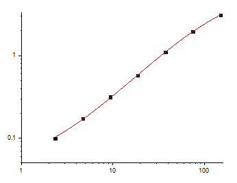
Dynamic range ~2.35 – 150 pg/mL (7-point standard curve)
Detection Colorimetric TMB → 450 nm read (620–650 nm reference optional)
Key components Pre-coated anti-IL-10 96-well plate · IL-10 protein standard · biotin-anti-IL-10 detection Ab · EliKine™ Streptavidin-HRP · standard/sample diluents · wash buffer · TMB substrate · stop solution · sealing films
Storage / Ship 2–8°C (unopened); ship blue-ice gel pack; For research use only
The competitive edge is boring in the best way: the plate is pre-coated, the standard is supplied, and the capture/detection antibody pair is pre-validated to give you specific IL-10 signal without the background creep that ruins low-end resolution. Translation—your 1–3 pg/mL neighborhood stops being a guessing game.
What Actually Improves When You Stop Winging the IL-10 Readout
① Your "non-significant" baseline samples become real numbers.
With a 1 pg/mL floor and a calibrated standard curve, you interpolate—rather than marking <LOD and hoping your stats package forgives you. For immunology reviewers, that is the difference between a defensible bar plot and a supplement they question.
② Treg / M2-polarized / tolerogenic DC panels stop looking like noise.
IL-10 is supposed to be the story there. But if your CV balloons because everyone coats or dilutes slightly differently, the effect size vanishes into error. The pre-coated format + lot-controlled reagents keep inter-assay CV in the respectable range.
③ Multi-cytokine stories land smoother.
Run TNF-α (KTE6015/KTE6006-type range), IL-6 (KTE6017), IL-1β (KTE6013), and IL-10 (KTE6019) from the same harvest—each on its own pre-coated plate, each with its own standard—so the ratio (pro- vs. anti-inflammatory) is built on comparable precision, not mixed-method heroics.
Bench-Level Rules That Keep Your 450 nm Curve From Betraying You
These come straight out of the operational notes shipped with the kit—and they're the exact lines reviewers wish every Methods section included:
• RT balance: Bring all components to room temperature (≥30 min) before opening. Cold reagent droplets on a warm plate lid = condensation = drift.
• Never cross-mix lots: Don't "borrow" wash buffer or Streptavidin-HRP from a different batch or brand. The standard curve is lot-calibrated.
• Fresh tips, every transfer: IL-10 is tiny, sticky, and one contaminated well cascades through the whole standard.
• Seal it back fast: Unused strips → back into the foil pouch with desiccant → 2–8°C. Moisture destroys pre-coated plates silently.
• Mix, but don't massacre: gentle orbital shake or a manual tap every ~10 min during incubations is the recommendation—no vortex-the-plate chaos.
• Duplicates/triplicates: Always. The kit notes explicitly recommend it; your CV will thank you.
Where KTE6019 Earns Its Spot in Real, Funded Work
Research Context Why IL-10 + This Format Is the Right Call
Sepsis immunosuppression / "compensatory anti-inflammatory response syndrome" (CARS) Serum IL-10 surges predict anergy and secondary infection risk; 1 pg/mL sensitivity captures the early trajectory
Treg / Tr1 / tolerogenic DC / transplant tolerance models Low-baseline, stimulus-dependent secretion—needs a tight curve, not a "positive/negative" call
Tumor microenvironment & immune evasion Myeloid-derived suppressors and Tregs hide behind IL-10; quantifying it cleanly strengthens "immune exclusion" arguments
Neuroinflammation / microglia polarization Glia-secreted IL-10 is part of the repair signature; supernatant volumes are often limiting → pre-coated microplate protects material
Parasitology / helminth-immune modulation Classic IL-10–dominant Th2/tolerance phenotypes; cross-species antigen prep makes reagent consistency even more critical
A Clean Methods Paragraph You Can Borrow
Human IL-10 was quantified using a quantitative sandwich ELISA (EliKine™ Human IL-10 ELISA Kit, KTE6019; Abbkine) according to the manufacturer's protocol. A microplate pre-coated with capture anti-IL-10 was incubated with standards and samples; following washing, a biotinylated anti-IL-10 detection antibody and Streptavidin-HRP were applied, developed with TMB, stopped, and read at 450 nm (620–650 nm reference). Values were interpolated from the supplied recombinant IL-10 standard curve and expressed as pg/mL (or normalized to total protein/cell number as indicated).
Explore the EliKine™ Human IL-10 ELISA Kit (KTE6019) full specs, manual & ordering options here:
🔗 https://www.abbkine.com/product/elikine-human-il-10-elisa-kit-kte6019/
(For research use only. Not for human or clinical diagnostic use.)





