


The Megadalton Slime That Predicts Liver Failure, Tumor Invasion, and Joint Destruction: Why Your HA Number Shouldn't Come From a Dye-Binding Hack — And How KTE61936 Actually Gets It Right
Hyaluronic acid (more precisely hyaluronan / hyaluronate, HA) is the only glycosaminoglycan in your body that isn't sulfated, isn't built in the Golgi, and can reach a molecular weight of 4,000–10,000+ kDa — a single polymer chain long enough to span the entire pericellular coat and physically gate which proteins, growth factors (HGF, FGF-2), and immune cells touch the cell surface. It's the frictional cushion in your synovial fluid (giving it that egg-white viscoelasticity that lets joints bear load without grinding), the hydration sponge in your dermis, the matrix organizer in liver sinusoids, and — critically for anyone running a cancer or fibrosis lab — a dynamic, actively turned-over signal whose blood levels track endothelial barrier breakdown, fibroblast activation, and the transition from compensated cirrhosis to decompensation. Yet most labs still measure it with the dignity of a carbazole color assay or a slot-blot that can't tell HA from any other uronic acid. The Human Hyaluronic Acid (HA) ELISA Kit (KTE61936) from Abbkine is the reagent that drags this megadalton GAG out of the dark ages and puts it on a quantitative, plate-readable curve (ng/mL) with the specificity and CVs a real dataset demands.
HA in One Paragraph: A 4,000-kDa Polymer That Acts Like a Molecular Gatekeeper
Structurally, HA is dead simple: a linear, nonsulfated repeating disaccharide — [→β-D-GlcA-(1→3)-β-D-GlcNAc-(1→4)→]ₙ — but its size is what makes it biologically unique. Synthesized by HA synthase enzymes (HAS1/2/3, HAS2 being the dominant one) at the inner leaflet of the plasma membrane (yes, plasma membrane, not the Golgi like every other GAG), HA is extruded directly into the ECM as a growing chain, where it:
HA Role Mechanism Why It Matters Clinically
ECM hydration & porosity Each disaccharide carries two carboxylates → binds Na⁺ and water electrostatically → creates the hydrated gel that spaces collagen and controls diffusive access Skin turgor, glomerular filtration barrier, liver sinusoidal fenestration
Receptor signaling (CD44, RHAMM/IHABP, TLR4) CD44 is the primary signaling receptor — HA fragments (oligo-HA, ~6–20 disaccharides, < 200 kDa) activate CD44 → ERK/AKT, Rac1, PI3K, NF-κB → pro-motility/pro-inflammatory RA synovium, cancer invasion fronts, inflammatory edema
Synovial lubrication High-MW HA (> 1,000 kDa) is the viscoelastic carrier in joint fluid; depleted/downshifted MW = arthritic friction Osteoarthritis progression
Liver sinusoidal barrier Fenestrated endothelium retains HA on its surface via CD44/HA-receptors; when the barrier erodes, serum HA spikes because the liver can no longer clear it Serum HA = functional marker of sinusoidal capillarization / fibrosis burden
The kicker: HA exists as a massive size continuum in vivo — from full-length megadalton chains (healthy, anti-angiogenic, CD44 adhesive) to depolymerized oligos (pro-angiogenic, pro-inflammatory, TLR4-active). Your assay needs to know which version it's seeing — or at least give you a total HA mass that correlates with what the tissue is shedding.
The Critical Caveat Every HA Paper Must State Up Front: It's Not a Protein — So What Does "Sandwich ELISA" Mean Here?
This is where 90% of the confusion lives. You cannot run a true two-epitope sandwich on a disaccharide repeat chain the way you do with IL-6 or Bax. What HA "ELISAs" actually deploy is a binding-protein–based immunoassay format:
The KTE61936 architecture uses purified HA-binding protein (HABP, derived from cartilage/link protein or recombinant CD44-derived domains) as the immobilized capture on the microplate. HABP binds HA through a defined structural pocket (not an "epitope" in the peptide sense, but a conformational glycan motif). Then:
- A biotinylated second HABP/detector conjugate binds a different accessible region of the same HA chain (allowing the "sandwich" logic to work because the polymer has multiple binding sites along its length).
- Streptavidin–HRP → TMB → color ∝ bound HA mass.
- 450 nm → interpolate from a human HA standard curve.
The consolidated specs from distributor/technical data aligned with KTE61936:
Parameter KTE61936-class Specification
Target Human HA / Hyaluronan / Hyaluronate (repeating [GlcA-GlcNAc]ₙ)
Format Sandwich-type immunoassay (HABP-capture / biotin-HABP detection) — colorimetric, 450 nm
Range 0.156 – 10 ng/mL (calibrated against human umbilical-cord HA standards)
Sensitivity / LOD ~0.039–0.047 ng/mL
Intra-Assay CV < 6–8%
Inter-Assay CV < 10%
Specificity No significant cross-reactivity with other GAGs (CS, DS, HS, KS) at physiological levels
Samples Serum, plasma, CSF, tissue homogenates, cell culture supernatants / lysates
Assay time ~3–5 hours
(Always confirm exact range, standard identity [umbilical vs. rooster comb], and lot-specific recovery on the shipped Abbkine datasheet.)
Sample Handling: HA Is a Chain — And Chains Snap If You're Rough
This is the single most under-discussed point in HA work: HA is a physical polymer. Vortexing serum like it's a PBS buffer, or freeze–thawing three times, will shear full-length HA into fragments that may or may not be recognized the same way by the HABP pair — and your "HA level" suddenly becomes an artifact of your pipette force.
Golden rules:
- Collect in EDTA or plain tubes (avoid heparin if possible — heparin is itself a GAG and some HABPs have weak heparan/HS cross-talk at high concentrations).
- Spin cold (4°C), aliquot, -80°C, single thaw — handle gently, no vortex on high.
- Minimize mechanical shearing: pipette with wide-bore tips for viscous samples; avoid repeated passage through narrow-bore needles.
- For tissue: homogenize in cold PBS + protease inhibitors, clarify gently, and run the supernatant. Report as ng HA / mg total protein (BCA) — not "ng/mL tissue" (meaningless without normalization).
Where HA Quantification Actually Carries the Paper
- Liver Fibrosis & Sinusoidal Capillarization (The Best-Validated Clinical Biomarker Story)
This is where HA earned its reputation. The liver sinusoidal endothelial cell (LSEC) fenestrae and the perisinusoidal CD44⁺ stellate-cell–associated matrix are the body's primary HA clearance sink — HA binds, is internalized, and degraded by HARE (HA receptor for endocytosis, also called Stabilin-2). As fibrosis progresses → fenestrations collapse → HA clearance ↓ → serum HA ↑, it becomes one of the few ECM markers that tracks functional barrier loss, not just collagen accumulation. In compensated vs. decompensated NASH/cirrhosis cohorts, HA often outperforms PIIINP or TIMP-1 for discriminating mid-stage burden, and it's routinely used alongside FIB-4 / elastography in multi-marker panels.
- Osteoarthritis & Synovial Fluid Dynamics
Healthy synovial HA = 1,000–4,000 kDa, ~1.4–3.6 mg/mL in the joint space — that's not a trace analyte, it's a structural lubricant. In OA, two things happen:
• Total HA concentration in SF drops (washout + reduced synovial fibroblast synthesis)
• Mean MW downshifts (ROS/HYAL1/HYAL2 depolymerization → oligo-HA fragments → CD44/TLR4 pro-inflammatory signaling)
Running KTE61936 on serum (easier, indirect) or CSF/aspirate (direct) gives you the mass readout; pairing it with HYAL activity or oligo-HA fragment analysis rounds out the story.
- Cancer Desmoplasia, Invasion Fronts & Lymphatic Spread
HA is a CD44 ligand, and high stromal HA + CD44v expression = classic poor-outcome signature in breast, colorectal, pancreatic, ovarian, and head-and-neck contexts. Tumors that saturate their stroma with HA create a hydration channel that lets cells migrate/intravasate more easily; conversely, HA oligos from stromal HYAL cleavage feed CD44 signaling loops that drive invasion. Quantifying total HA in tumor lysates/homogenates (ng/mg protein) is the ECM-burden anchor for any CD44/Wnt/HGF crosstalk figure.
- Inflammatory Lung Injury & ARDS (The Capillary Leak Axis)
Acute lung injury → endothelial glycocalyx + HA-rich pericellular coat shed into circulation → serum/plasma HA spikes sharply (often one of the earliest ECM markers to move). Tracking it alongside ANGPT2, vWF, and SP-D frames the endothelial-barrier-collapse narrative better than BAL albumin alone.
- Wound Healing, Skin Aging & Dermal Hydration
Dermal fibroblast HAS2 → HA fills the ground substance; UV/ROS downregulate HAS2 and upregulate HYAL → dermal HA ↓ → wrinkle + impaired healing. Measuring HA in punch-biopsied skin or suction-blister fluid (ELISA normalized to hydroxyproline or BCA) is the compositional readout that links cosmeceutical or UV-protection studies to matrix content, not just a "TEWL or moisture meter" device reading.
- CRISPR/Overexpression Validation (HAS2 / HYAL1-2 / STAB2)
Editing HAS2 (↑HA), HYAL1/2 (↓HA MW), or STAB2 (↓HA clearance) → report HA mass ± SEM from the calibrated curve (ng/mg), not a toluidine-blue spot. Co-label with CD44 IF, p-AKT/p-ERK, and Ki-67 so the matrix change has its receptor-linked phenotype.
Quick Practical Notes for the Plate
• Dilute into kit buffer per the manual (HA at pg–ng/mL is adsorptive; low-binding plates/plastic help but the kit's formatted wells already carry the capture HABP).
• Warm all reagents ≥ 30 min RT before opening; protect TMB from light; stop uniformly; read 450 nm promptly.
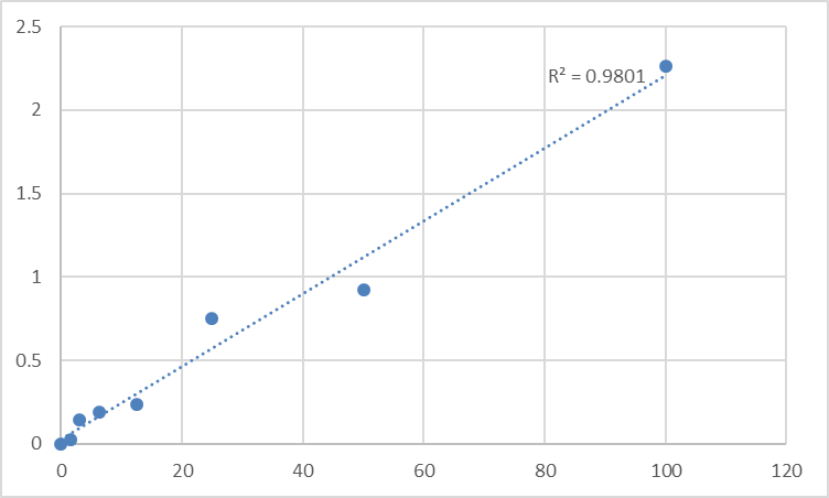
• Fit a 4-PL to the standard curve (HA immunoassays are curved, not linear at the knees).
• Run the full standard curve on every plate — GAG adsorption variability is exactly why plate-to-plate normalization needs the curve.
The Bottom Line
HA is the megadalton, nonsulfated GAG that hydrates your dermis, cushions your joints, gates growth-factor access, and — when its liver clearance collapses — screams "fibrosis" louder than any collagen stain. It's not a protein, so it can't be measured like one; it needs a binding-protein–based sandwich immunoassay that respects its polymeric nature and handles it gently enough to keep the chain intact. The Human Hyaluronic Acid (HA) ELISA Kit — KTE61936 from Abbkine is built for exactly that: HABP-capture → biotin-HABP detection → HRP–TMB → 450 nm → ng/mL, over a 0.156–10 ng/mL calibrated range with LOD ~0.04 ng/mL, in a ~3–5 hour workflow that scales from a cirrhotic-serum bank to a synovial-fluid cohort without a carbazole melting block in sight.
Product Reference: KTE61936 – Human Hyaluronic Acid (HA) ELISA Kit
Learn more and order: https://www.abbkine.com/product/human-hyaluronic-acid-ha-elisa-kit-kte61936/
(For Research Use Only; not for diagnostic procedures in humans.)


