




The Far-Red Deficit: Why Your In Vivo Imaging Has Been Broadcasting Autofluorescence Instead of Cell Data—And How DiD's Spectral Shift Finally Silences the Background That Plagued Membrane Tracing for a Generation
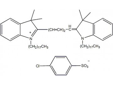
Every researcher tracking labeled cells in a living animal knows the reckoning: the abdominal region glows identically whether cells are injected or not. Liver flavins and dietary porphyrins autofluoresce exactly where DiI emits. You are not imaging your cells; you are imaging the mouse's last meal over hepatic noise. This is not a failure of the carbocyanine family—DiI and DiO have served decades admirably in cultured cells and thin sections. Their limitation is physical: biological tissue absorbs visible light fiercely, and endogenous fluorophores flood the 400–600 nm range, frequently exceeding specific signal. Shifting to the far-red/near-infrared, beyond ~630 nm, slashes autofluorescence by orders of magnitude and extends penetration from micrometers to millimeters. Abbkine's DiD (DiIC18(5)), Catalog No. BMD0073, is the deep-red solution: CAS 362596-00-7, molecular formula C₆₇H₁₀₃ClN₂O₃S, molecular weight 1052.1, a dark blue solid emitting far-red (λEx/λEm: 644/663 nm in methanol). Its spectral positioning transforms whole-animal fluorescence imaging from qualitative visualization into quantitative analysis, giving stable, long-term membrane labeling that visible-wavelength dyes simply cannot provide in complex tissues.
BMD0073's Optical Identity: The Spectral Shift That Converts Tissue Autofluorescence into Irrelevant Background
The excitation/emission maxima—644/663 nm—place BMD0073 well beyond the autofluorescence range of flavins, porphyrins, and lipofuscin. This is not a convenience; it is the biochemical foundation of in vivo cell tracking. This lipophilic carbocyanine is weakly fluorescent in water but highly fluorescent and quite photostable when incorporated into membranes, with an extremely high extinction coefficient and short excited-state lifetimes of ~1 nanosecond in lipid environments. Once applied, the dye diffuses laterally, staining the entire cell surface uniformly. The high extinction coefficient ensures efficient photon capture; the short lifetime minimizes the window for oxidative degradation. DiD can be excited by the 633 nm He-Ne laser and has longer wavelengths than DiI, which is critical in specimens with significant intrinsic fluorescence. At 565 nm (DiI emission), liver and spleen autofluorescence can overwhelm specific signal fivefold. At 663 nm (DiD emission), those same fluorophores' extinction coefficients drop by one to two orders of magnitude, transforming marginally interpretable images into quantitatively analyzable data. For tracking CAR-T cells in spleen and liver—organs nearly opaque to DiI yet substantially transparent at DiD wavelengths—this spectral separation is not an advantage; it is the prerequisite for collecting any interpretable data.
The Protocol Where Most Far-Red Dyes Fail—And Where BMD0073 Delivers Uniform, Stable Fluorescence
BMD0073 labels membranes with uniformity and stability that generic far-red dyes cannot match. The dye inserts into the membrane and diffuses rapidly, staining the entire surface evenly. Critically, it exhibits high lateral mobility but does not transfer between adjacent cells under normal culture conditions—a property called stable integration, avoiding the false-positive bystander labeling caused by rapid internalization or leakage seen with many other membrane dyes. For lineage tracing, extravasation monitoring, and transplantation tracking, this eliminates the ambiguity that confounds migration studies when fluorescent labels exchange between populations. Staining is straightforward: load via simple incubation in serum-free or low-serum medium for 5–20 minutes at 37°C, followed by a brief wash. The dye is nontoxic at recommended 1–5 μM concentrations, and labeled cells remain viable for flow cytometry, microscopy, and in vivo transplantation. Based on 100 μL of a 5 μM working solution per staining, 10 mg of product enables approximately 19,009 reactions—a volumetric yield supporting large-scale studies. Post-staining, paraformaldehyde fixation is compatible; methanol or other organic solvents must be avoided as they extract the lipophilic dye. Intriguingly, plasma membrane staining also performs well after fixed permeabilization with 0.1% Triton X-100 at room temperature, accommodating protocols where intracellular antigen detection precedes membrane labeling. The far-red emission (collected in the APC/Cy5 channel) leaves the entire visible spectrum—blue, green, orange, red—available for other fluorophores, enabling four-color panels without spectral overlap penalties.
In Vivo and Ex Vivo Performance: The Tissue Contexts Where BMD0073 Transforms Marginal Signal into Quantitative Data
BMD0073's practical advantage crystallizes when imaging labeled cells in tissue contexts that defeat visible-wavelength dyes. The far-red emission shifts detection into a window where autofluorescence drops by orders of magnitude, enabling deep-tissue imaging. Researchers have used DiD to track mesenchymal stem cells in a rat knee osteoarthritis model with no cytotoxicity, visualizing populations at depths inaccessible to DiI or DiO. Peer-reviewed and technical data document successful tracking of stem cell migration to injury sites, monitoring of CAR-T cell persistence in xenografts, and visualization of neuronal projections in organotypic slice cultures. The dye has even been employed for precise 3D localization of intracerebral implants using a simple brain clearing method, revealing electrode localization in transgenic mice expressing cell-specific fluorescent markers. Long-term stability is the second critical parameter: labeled cells can be tracked for extended periods with no dye leakage and no transfer to host cells. For CAR-T therapy studies monitoring therapeutic cell persistence over weeks, this membrane retention without intercellular transfer makes the experiment interpretable.
Three Publications, Journal of Hazardous Materials (IF 26.6), and Peer-Reviewed Validation in the Biomedical Literature
A fluorescent dye's credibility rests on independent peer-reviewed data. BMD0073 has been cited in 3 publications. The most prominent is a study in the Journal of Hazardous Materials (IF 26.6) on engineered outer membrane vesicles for enhancing solid tumor CAR-T cell therapy—a context requiring dye that withstands vesicle purification, CAR-T staining, and in vivo imaging without degradation or transfer. The Chinese-language product page records 1,656 views, reflecting sustained interest in a far-red membrane dye for deep-tissue imaging. Additional citations are available through the product page, and while the CiteAb entry currently reports zero open-access mentions, the growing body of published work validates BMD0073 across immunology, oncology, and neuroscience. For researchers transitioning from visible-wavelength dyes, this record provides community-tested evidence in contexts where spectral separation from autofluorescence determines whether data are interpretable. Note the alternative catalog designation BMD00073; researchers should verify the correct number (BMD0073) when ordering.
Practical Protocol Decisions That Distinguish Publication-Grade Far-Red Labeling
DiD is highly hydrophobic and must first be dissolved in an organic solvent such as ethanol or DMSO before aqueous dilution; direct addition to buffer causes aggregation and punctate background. Store at -20°C protected from light for up to 12 months; while stable at 4°C for months, avoid freeze-thaw cycles. Aliquot DMSO stocks into single-use volumes to prevent degradation. Paraformaldehyde fixation is compatible; avoid methanol and acetone. If Triton X-100 permeabilization is required, perform it before staining, as DiD works well after fixed permeabilization. For flow cytometry, collect fluorescence in the APC channel (excitation: 633–640 nm; emission: 660/20). Because DiD occupies an underutilized far-red channel, adding a membrane label typically does not require panel redesign. This product is for research use only, not for diagnostic procedures.
The Abbkine Carbocyanine Ecosystem: A Complete Spectral Palette
BMD0073 occupies the far-red channel (644/663 nm) within a family spanning DiO (green, 484/501 nm, BMD0072), DiI (orange-red, 549/565 nm, BMD0071), and DiR (near-infrared, BMD0074). This palette enables multiplexed membrane labeling from blue to NIR. Two populations can be distinguished with DiO and DiI; three with the addition of DiD; four with DiR. All share the same C18 alkyl chain chemistry, environment-dependent fluorescence, and protocol logic—reducing technical overhead and ensuring that adding a far-red channel doesn't introduce incompatible chemistry. The consistent quality control framework—HPLC purity, fluorescence validation, lot-to-lot consistency—extends across the portfolio.
Product Details:
- Product Name: DiD (DiIC18(5))
- Brand: Abbkine
- Catalog Number: BMD0073
- Alternative Designations: BMD00073
- CAS Number: 362596-00-7
- Molecular Formula: C₆₇H₁₀₃ClN₂O₃S
- Molecular Weight: 1052.1
- Formulation: Dark blue solid; soluble in DMSO or ethanol
- Excitation/Emission: λEx/λEm: 644/663 nm (MeOH); far-red fluorescence
- Features & Benefits: High extinction coefficient; short excited-state lifetimes (~1 ns) in lipid environments; highly fluorescent and quite photostable when incorporated into membranes; weak fluorescence in water; longer wavelengths than DiI; compatible with 633 nm He-Ne laser; stable integration with no intercellular transfer; excellent signal-to-noise due to reduced autofluorescence
- Applications: Cell membrane labeling; long-term cell tracking; neuronal tracing; in vivo cell migration and biodistribution; whole-animal fluorescence imaging; multiplexed panels (far-red channel); flow cytometry; stem cell homing studies; CAR-T cell persistence monitoring; organotypic slice culture; 3D implant localization; cytotoxicity assays; lipoprotein labeling
- Working Concentration: Typically 1–5 μM; 10 mg sufficient for ~19,009 staining reactions at 5 μM in 100 μL
- Storage: -20°C, protect from light; stable for up to 12 months
- Shipping: Gel pack with blue ice
- Citations: 3 peer-reviewed publications
Product Link: https://www.abbkine.com/product/did-diic185-bmd0073/





