


The 286-Da Fat-Soluble Ghost in Your Mouse Chow: Why "Vitamin A Status" Is Never Just a Diet Label — And How the Competitive Immunoassay Behind KTE70032 Finally Gives You Retinol Numbers That Survive Peer Review
Vitamin A has arguably the worst reputation-to-actual-molecular-size ratio of any "vitamin" in the mouse house. Everyone knows the headline — night blindness, xerophthalmia, immune collapse — but almost nobody treats VA / all-trans-retinol (C₂₀H₃₀O, MW 286.45 Da) like the fat-soluble, light-labile, protein-binding small molecule it actually is. Retinol doesn't float freely in aqueous plasma; it rides in a triangular chaperone system: retinol-binding protein 4 (RBP4, 23 kDa) + transthyretin/TTR (55 kDa dimer) in serum, and when it enters tissue, it swaps onto cellular retinol-binding proteins (CRBP-I/II) before being oxidized to retinaldehyde and ultimately to all-trans-retinoic acid (atRA) — the ~300 Da ligand that actually binds RAR/RXR nuclear receptors and reshapes transcription. The Mouse Vitamin A (VA) ELISA Kit (KTE70032) from Abbkine exists because you can't run an HPLC-fluorescence reference method on 96 samples before your postdoc quits, and you definitely can't pretend a 286-Da hapten is a sandwich-ELISA protein. It's a competitive hapten immunoassay — VA–protein conjugate coated, sample retinol competes with a labeled detection reagent, signal runs downhill, and you interpolate pg–ng/mL from a 4-PL curve — so your dietary-restriction, liver-stellate, or neuro-developmental paper reports an actual retinol concentration, not "the diet was VA− so it must be low."
VA / Retinol in One Paragraph: The ~286-Da Diterpenoid That Controls Vision, Tolerance, and Lineage Choice
Chemically, "Vitamin A" in the nutritional sense is the alcohol form = all-trans-retinol (or 11-cis-retinal in the eye) — a β-ionone ring + isoprenoid polyene tail + primary alcohol, absorbing strongly at 325 nm (that pale yellow tint of old liver oil is literally π→π* transition density). The metabolic sequence everyone memorizes:
Step Enzyme / Carrier Product Function
Dietary → intestinal absorp. Pancreatic lipase / bile salt micelles → taken up as retinyl esters → LRAT (lecithin:retinol acyltransferase) re-esterifies in enterocyte → chylomicrons → lymph → liver Retinyl palmitate / stearate (storage ester) Liver stores > 80–90% of total body VA as stellate-cell lipid-droplet retinyl esters
Mobilization (fasting/stress) HRP (hepatic retinyl ester hydrolase / acyl-CoA:retinol O-acyltransferase reverse) → retinol released → binds RBP4 → TTR stabilizes → circulates Retinol–RBP4–TTR complex (~80–100 kDa) The only way VA safely travels an aqueous compartment
Tissue uptake STRA6 (RBP4 receptor) / LRAT in target cells Retinol → retinal (RDH) → retinoic acid (Raldh1/2) atRA = RAR/RXR ligand → Hox patterning, thymic tolerance, neurogenesis, germline maintenance
The clinical mouse number that matters: serum retinol in a well-fed C57BL/6 is roughly 1–2 μM (~280–570 ng/mL), but the dietary VA− / VAD (vitamin A-deficient) model drives it down to < 0.1–0.2 μM (< 30–60 ng/mL) over 6–12 weeks — and that drop propagates through thymic involution, defective Treg generation, impaired mucosal barrier immunity, and defective spermatogenesis long before anyone sees a weight change.
Why a Competitive Hapten Immunoassay — And Why "Sandwich" on a 286-Da Molecule Is a Category Error
Retinol has one ionizable OH, no peptide backbone, and a surface area roughly the size of a large amino acid — there is categorically no room for two spatially independent, non-overlapping epitopes for a classical sandwich. What happens instead is the textbook competitive ELISA / hapten format:
The Architecture Inside KTE70032
Variant implementations float across vendor docs, but the canonical competitive principle works one of two symmetric ways:
Route A (antigen-coated, most common):
- Microplate is coated with VA–protein conjugate (VA–BSA or VA–OVA) — the immobilized hapten-carrier acts as the "capture surface."
- Sample VA + a fixed, limited amount of detection reagent (e.g., anti-VA antibody → biotinylated → SA–HRP, or HRP-labeled anti-VA detection conjugate) are added together → they compete for binding sites.
- Wash → TMB → 450 nm → signal INVERSELY ∝ [retinol]: more retinol in your sample outcompetes the plate-bound VA → lower OD.
Route B (antibody-coated):
- Anti-VA pAb/mAb pre-coated
- Add sample VA + HRP-/biotin-tracer (VA–enzyme conjugate) → competition → same inverse readout.
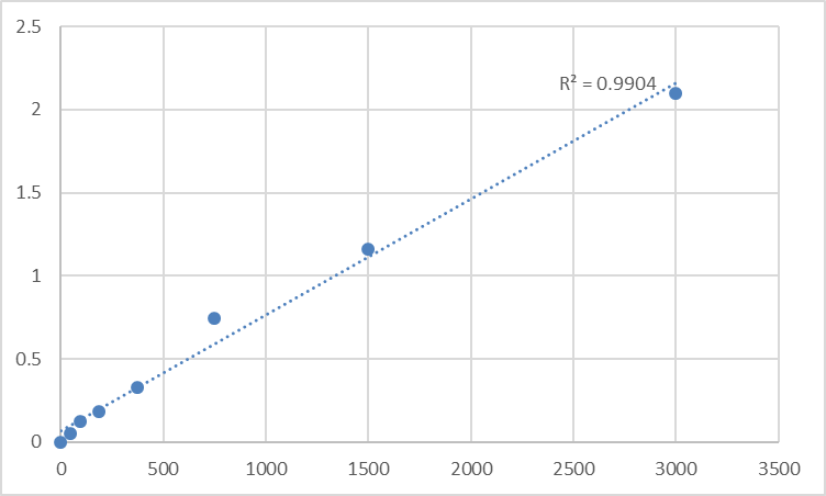
Either way, the governing relationship is Bound Tracer ∝ 1/[Free VA], fitted as a 4-parameter logistic (4-PL) or log-logit (B/B₀) curve — never linear.
Consolidated performance envelope from the distributor/technical mirrors aligned with this kit class (range varies by standard identity/units; ng/mL vs pg/mL are trivially interconvertible × 286):
Parameter KTE70032-class Specification (competitive hapten immunoassay)
Target Mouse Vitamin A / VA / all-trans-retinol (C₂₀H₃₀O, MW 286.45)
Assay Type Competitive ELISA (hapten format — not a true sandwich; some vendor sheets lazily shorthand it "sandwich/夹心法" but the stoichiometry is competition)
Detection Biotin-Ab / HRP–tracer → TMB, read 450 nm (inverse curve)
Dynamic Range Commonly 0.156 – 10 ng/mL (≡ 156 – 10,000 pg/mL), with alternate calibrations reported as 187.5 – 3,000 pg/mL depending on standard
Sensitivity / LOD 0.094 – 0.156 ng/mL (94–156 pg/mL); select formats quote ~9.4 ng/mL LOD when the standard is calibrated in higher-range units — always confirm your lot CoA
Intra-Assay CV < 8–10%
Inter-Assay CV < 10–12%
Specificity Optimized for VA/retinol; minimal cross-reactivity with unrelated carotenoids when the antibody is well-raised (β-carotene, lutein, zeaxanthin, α-tocopherol are structurally distinct enough to reject at physiological levels in clean extracts)
Samples Serum, plasma (EDTA/heparin), tissue homogenates (requires organic extraction step), cell culture supernatants, other biological fluids
Assay time ~2.5–4 hours
(Confirm exact range, standard identity/traceability, and dilution scheme on the shipped Abbkine datasheet/CoA for KTE70032.)
The Sample-Prep Rule That Separates "Retinol" From "Chlorophyll-Colored Chaos"
Because retinol is fat-soluble, light-sensitive, and largely RBP4-bound, the prep is half the science:
Serum / Plasma (the cleanest matrix)
• Collect in EDTA or heparin (not citrate if you can avoid it), wrap tube in aluminum foil or keep dark, keep on ice, spin ≥ 2,000 ×g, 10 min, 4°C within 30–60 min, aliquot into amber low-bind tubes, snap -80°C, avoid >1 freeze–thaw.
• Many labs do a single-step deproteinization/extraction (ethanol + hexane or petroleum ether, brief vortex, spin, collect organic phase, evaporate N₂, reconstitute in kit buffer) to strip RBP4 and lipids that could blur the hapten–antibody equilibrium — follow the manual's exact extraction instruction; if it says "no extraction needed for serum," it means the antibody tolerates the protein matrix at the recommended dilution.
Tissue (liver, lung, brain, fat)
• Snap-frozen tissue → homogenize in ice-cold PBS + protease inhibitors, then add 1–2 volumes ethanol + an equal volume of hexane/pet-ether, vortex, spin, collect the organic (upper) phase (contains retinol + other carotenoids) → dry under N₂ stream → reconstitute in kit assay buffer → read.
• Express as ng retinol / g tissue wet weight or ng / mg total protein (BCA on the aqueous pellet).
Golden rule: protect from light. Retinol photo-oxidizes to retinal + retinoic acid + epoxides under fluorescent white light in ~20–60 min. If your samples turn yellow in the tube, your number is drifting.
Where Mouse VA/Retinol Quantification Actually Carries the Paper
- Dietary VA Deficiency (VAD) & Immune Reconstitution — The Thymus–Treg Axis
This is the flagship. VAD → thymic epithelial cell (TEC) retinoic-acid signaling drops → AIRE expression dips → central tolerance weakens → autoreactive clones escape → peripheral Treg generation (Raldh2⁺ DCs → atRA → Foxp3 induction) collapses. The measurable endpoint that ties diet to thymus is serum retinol (ng/mL) + hepatic retinyl ester store (ng/g) — and the competitive ELISA is what lets you run basal → wk 4 → wk 8 → wk 12 on 40–60 EDTA-plasma samples and prove the drop was real, not just "we fed VA− chow."
- Liver Stellate Cell Biology & Fibrosis (The Retinoid Drop as a Scar Surrogate)
Quiescent HSCs (hepatic stellate cells) are the body's VA warehouse — they hold > 80% of total body retinyl esters in lipid droplets. Activation (any injury: CCl₄, BDL, NASH diet) → HSCs lose those droplets → retinol/retinyl ester in liver tissue ↓↓, while serum retinol may stay surprisingly stable (RBP4 release compensates until late failure). The power move: measure liver VA by ELISA on the organic extract (ng/g) alongside Desmin/GFAP (HSC activation) + α-SMA + collagen I/III — the falling-retinoid-in-tissue is the HSC-activation clock.
- Neurodevelopment, Visual Cycling & the "Brain VA" Question
Retinoids in the embryo / neonatal brain pattern the hindbrain, spinal cord, and retina (Hox genes, neural crest, cornea-limbal stem niche). Measuring brain/retina retinol + atRA equivalents (often ELISA for total VA + LC-MS for the active fraction) across VAD dams vs. pair-fed controls is the gold-standard nutritional-epigenetic model — and the ELISA gives you the high-throughput first-pass so you only send the informative samples to LC-MS.
- Gut Barrier & Mucosal IgA: The SIgA–Retinoid Cross-Talk
Vitamin A is the prerequisite for intestinal DC Raldh2 → atRA → gut-homing α₄β₇ imprinting + Treg generation + sIgA class-switch. The canonical weanling VAD model (C57BL/6 pups weaned onto VA− vs. VA+ from P21–P42) uses serum retinol ELISA + mesenteric LN Foxp3⁺ % + sIgA in fecal pellets as the readout trio — and it's the oldest, cleanest nutritional-immunology paradigm in the textbook.
- Pharmacology / Drug-Indi Trial QA: "Does My Compound Interfere with Retinoid Homeostasis?"
Isotretinoin/acitretin (systemic atRA analogues), bexarotene (RXR-selective), even ethanol (interferes with retinol → retinal dehydrogenase flux) — all mess with the VA–retinoic acid axis. Running KTE70032 on terminal bleed EDTA plasma (control vs. drug arm) is the 45-minute QA that tells you whether your compound's "immune effect" might be riding a retinoid-shift confounder — especially in cancer/dermatology models where atRA signalling is the mechanism of interest.
- Zebrafish / Amphibian Cross-Over Work (When You Want Mammalian-Readout Precision)
Even non-mouse labs doing Xenopus / Danio retinoid-teratogenesis screens sometimes validate with a mammalian anti-VA immunoassay on extracted yolk-sac or embryo homogenates — the hapten format ports surprisingly well as long as the organic extraction is respected.
A Minimal Protocol Skeleton You Can Paste Into Methods
- Blood: collect in EDTA, wrap in foil, keep on ice, spin ≥ 2,000 ×g, 10 min, 4°C within 60 min, aliquot into amber tubes, snap -80°C, one thaw only.
- For serum: same foil-protection rule; clot 30 min at 4°C, spin, collect supernatant, foil-wrap, freeze.
- Tissue: homogenize frozen tissue in cold PBS → add ethanol + hexane → vortex → spin → collect organic phase → N₂ dry → reconstitute in kit buffer → BCA the leftover aqueous pellet if you want ng VA / mg protein.
- Warm reagents ≥ 30 min RT before opening (but keep tracer/standards protected from light); read 450 nm promptly after stop; fit 4-PL / log-logit (B/B₀); run full standard curve per plate — competitive curves need that descending anchor, never skip B₀.
The Bottom Line
Vitamin A is the ~286-Da fat-soluble diterpenoid all-trans-retinol that the liver warehouses as stellate-cell retinyl esters, shuttles through the blood as an RBP4–TTR complex, and converts locally into retinoic acid to drive every RAR/RXR decision from thymic tolerance to Hox patterning — which means it's too small for a sandwich, too light-sensitive for a lazy cold chain, and too bound-to-protein for a "just spin and read" assumption. Measured correctly, it lives in a competitive hapten immunoassay whose inverse OD₄₅₀ → 4-PL curve gives you pg–ng/mL retinol you can batch, normalize, and defend. The Mouse Vitamin A (VA) ELISA Kit — KTE70032 from Abbkine runs that format: VA–protein conjugate coated (or anti-VA coated) → sample retinol competes with labeled detection → HRP–TMB → 450 nm → ng/mL interpolated, over roughly a 0.156–10 ng/mL calibrated envelope with LOD ~0.1–0.15 ng/mL, in a ~2.5–4 h workflow that scales from a VAD-thymus cohort to a liver-stellate timecourse without chaining you to an HPLC injection queue.
Product Reference: KTE70032 – Mouse Vitamin A (VA) ELISA Kit
Learn more and order: https://www.abbkine.com/product/mouse-vitamin-a-va-elisa-kit-kte70032/
(For Research Use Only; not for diagnostic procedures in humans.)


