


The 128-AA Trojan Horse: Why the Ubiquitin–Ribosome Fusion Protein UBA52 Is the Only "Dual-Use" Gene Your Proteostasis Paper Is Ignoring — And How KTE62310 Puts a Number on the Fusion
If your lab studies the ubiquitin–proteasome system, you spend most of your time staring at polyubiquitin chains, E1/E2/E3 cascades, and 26S proteasome activity assays — which is exactly why you're probably blind to the quietest, most evolutionarily clever source of cellular ubiquitin: not the polyubiquitin head-to-tail polymers (UBB/UBC genes), but a single ubiquitin moiety genetically welded to the N-terminus of a 60S ribosomal protein. That gene is UBA52 — official name Ubiquitin A-52 residue ribosomal protein fusion product 1, aliases CEP52, RPL40, HUBCEP52 (HGNC: 12458, Gene ID: 7311, Chr 19p13.11, UniProt: P62987) — and its protein product is the Ub–RPL40 fusion (~221 aa, computed ~22.8 kDa) that the cell transcribes, translates, and then precision-cleaves into two functionally opposite payloads: free monoubiquitin (76 aa, ~8.5 kDa) to feed the degradation pool, and mature ribosomal protein L40 (eL40) to lock into the 60S subunit. The Human Ubiquitin-60S ribosomal protein L40 (UBA52) ELISA Kit (KTE62310) from Abbkine exists to measure this dual-purpose precursor as a calibrated sandwich-ELISA signal (ng/mL) — so your proteostasis, ribosome-biogenesis, or cancer-autophagy story finally accounts for the protein that is simultaneously a ubiquitin donor and a ribosomal parts supplier, not just "another band at 22 kDa."
UBA52 in One Paragraph: Evolution's Two-For-One Deal
The human genome solves a logistics problem with elegant brute force. Cells need vast amounts of free ubiquitin (for conjugation) and vast amounts of ribosomal proteins (for translation). Rather than relying solely on independent transcription of a Ub gene + a separate RPL gene, evolution hard-wired two fusion genes — UBB (Ub–RPS27A) and UBA52 (Ub–RPL40) — where a functional Ub monomer is literally fused to the N-terminus of a core ribosomal protein via a short basic linker:
NH₂–[Ubiquitin 76 aa]–Gly-Gly–[linker ~2–20 aa]–[RPL40 / eL40 ~128 aa]–COOH
The life cycle is:
Step Enzyme / Machinery Outcome
Transcription / translation Pol II → ribosome Ub–RPL40 precursor (~22–23 kDa) appears first as an intact fusion in the cytoplasm
Processing (cleavage) DUBs (primarily USP9X/FAM/USP4 family at the linker) Ub released → enters free ubiquitin pool → fuels E1/E2/E3 cascade; RPL40 → incorporated into 60S subunit (cytoplasmic translation machinery)
Fusion that escapes cleavage Minor fraction remains intact Ub–RPL40 still attached → can be incorporated into ribosomes as the fusion (documented) and the Ub moiety can even be used for ribosome-associated ubiquitination (ribophagy regulation, RNF168-like histone crosstalk)
This is why UBA52 is ubiquitous (RefSeq expression: top-decile in ovary, bone marrow, and essentially every proliferating cell) — it's not a stress-induced luxury; it's a housekeeping logistics gene whose expression level tunes both how much ubiquitin the cytosol can summon and how fast the ribosome factory stocks its own shelves.
Why a Sandwich ELISA for a ~23-kDa Fusion — And Why "Anti-Ub Western" Is the Wrong Answer
Here's the trap 90% of labs walk into: they probe for UBA52 with anti-ubiquitin antibodies, see a ~8.5 kDa band and a ~22 kDa band, and say "Ub is there." But anti-Ub antibodies cannot tell you whether that Ub came from UBA52, from free monoubiquitin, from poly-Ub chains, or from one of the ~50+ UBB/UBA processed pseudogenes floating in the genome. The ~22 kDa band might be:
• Intact Ub–RPL40 fusion (what you want to measure)
• A cross-reactive Ub–RPS27A (UBB) or processed derivative
• Non-specifically retained protein in the gel front
A proper UBA52 ELISA solves this with capture + detection antibodies raised against the RPL40 C-terminal/linker region (or the Ub–RPL40 junction epitope) — not just the Ub moiety — so the readout is UBA52-specific, not "anything with a Ub fold."
The KTE62310 kit uses the field-standard architecture:
- Microplate pre-coated with a capture antibody specific for human UBA52 (directed at the fusion/junctional/RPL40 epitope, avoiding the Ub cross-reactivity swamp).
- Standards (recombinant UBA52) + samples — serum, plasma, tissue homogenates, cell lysates, cell culture supernatants/lysates, other biological fluids — added → UBA52 (intact fusion or accessible epitope pool) binds.
- Wash → biotinylated anti-UBA52 detection (different epitope) → Streptavidin–HRP → TMB → color ∝ bound UBA52.
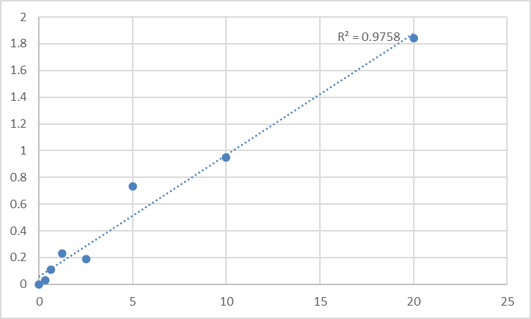
- Stop → 450 nm → interpolate ng/mL from the standard curve.
Consolidated performance envelope (from the search-aligned distributor/technical references for this kit family):
Parameter KTE62310-class Specification
Target Human UBA52 / Ub–RPL40 fusion (UniProt P62987, Gene 7311)
Format 96-well sandwich ELISA, pre-coated capture
Detection Biotin-Ab → SA-HRP → TMB, 450 nm
Dynamic Range 0.312 – 20 ng/mL
Sensitivity / LOD ~0.112–0.16 ng/mL
Intra-Assay CV < 8%
Inter-Assay CV < 10–12%
Specificity No significant cross-reactivity with UBB (Ub–RPS27A), free monoubiquitin, or other CEP-family members at physiological levels
Samples Serum, plasma (EDTA), tissue homogenates, cell lysates, culture supernatants
Assay time ~3–5 hours
(Confirm exact dilutions, recovery matrices, and the epitope strategy on the shipped Abbkine datasheet/CoA for KTE62310.)
Where UBA52 Quantification Actually Advances the Paper
- Proteostasis & the "Ubiquitin-Replenishment Budget"
This is the conceptual home. The free ubiquitin pool (~50–200 µM in cytosol) is maintained partly by proteasomal deubiquitination (DUBs releasing Ub from substrates) and partly by de novo synthesis from UBB and UBA52 precursors. When a cell is under proteotoxic stress (MG132, bortezomib, proteasome impairment, heat shock), the free-Ub pool drops — and UBA52 expression often compensates (it's one of the few Ub sources that can be transcriptionally ramped). Measuring UBA52 protein (ng/mg, BCA) during a CHX-pulse, MG132 washout, or HSF1/Nrf1 activation gives you the supply-side variable of the ubiquitin economy — the number that says "the cell is restocking Ub," not just "proteins are accumulating."
- HCC & Tumor-Autophagy: Inhibition of UBA52 → EMC6 → Tumor Suppression
A beautifully crisp recent finding (Zheng et al., Oncotarget/related journals): Inhibition/silencing of UBA52 induces autophagy via EMC6 and suppresses HCC tumorigenesis and progression. The mechanism threads through the RPL40–MDM2–p53 axis and broad ribosomal-output control. Here, UBA52 isn't "housekeeping" — it's oncogenically leveraged: the fusion maintains both Ub supply (protecting oncoproteins from destruction) and ribosome output (keeping the cancer cell's translation engine online). Quantifying UBA52 in HCC vs. adjacent nontumor lysates (ng/mg, normalized to actin or total protein, or to a ribosomal marker like RPL5/RPL7) is the supply-side readout that "proliferation markers went up" alone can't explain.
- CRC & lncRNA Crosstalk: LUCAT1 → UBA52 → RPL40–MDM2–p53
Colorectal models show lncRNA LUCAT1 binds UBA52 → stabilizes the Ub–RPL40 pool → RPL40 promotes MDM2-mediated p53 suppression → cell-cycle progression / survival. It's a striking reminder that the "ribosomal protein" isn't passive furniture: RPL40 is an MDM2-binding partner that modulates the guardian-of-the-genome. If your CRC line overexpresses an lncRNA or miRNA, report UBA52 protein remaining ± SEM from the calibrated curve — it's the molecular inventory that closes the loop between "lncRNA was up" and "p53 stayed silent."
- Gastric Cancer & the SMYD5–rpL40 Methylation Axis
SMYD5 methylates rpL40 (the RPL40 moiety of UBA52) — a histone methyltransferase family enzyme acting on a ribosomal substrate — and that methylation links ribosomal output tuning to gastric cancer phenotype. The fusion protein becomes the substrate. Measuring UBA52 (total fusion) alongside modified-rpL40 readouts or ribosome profiling ties the methylation to the actual scaffold it modifies.
- DNA Repair & Chromatin: UBA52/UBA80 Fine-Tuning RNF168-Dependent Histone Ubiquitination
This is the frontier: UBA80 (Ub–RPS27A) and UBA52 don't just feed free Ub — their Ub moieties can be presented in ribosomal or ribosome-proximal contexts, and recent work shows they fine-tune RNF168-dependent H2A/H2AX ubiquitination (the DSB-response histone mark). Translation-coupled ubiquitin availability becomes a chromatin-repair variable. Quantifying UBA52 in nuclear vs. cytoplasmic fractions (fractionation → BCA → ELISA) is the clean way to ask: did the repair defect trace to the Ub pool, or to the E3?
- HIV-1 & Retroviral Gag Incorporation
One of the more esoteric but cool facts: biotinylated Ub–RPL40 (UBA52) is incorporated into HIV-1 Gag virus-like particles — meaning the virus hijacks the host's Ub fusion machinery (or its cleavage products) as a structural co-opt. Not a mainstream assay for most labs, but a reminder that UBA52 is not a static storage protein — it's metabolically active, processed, and trafficked.
- CRISPR / shRNA Validation
Editing UBA52 is technically tricky (essential gene, Chr 19 housekeeping) — most labs use inducible shRNA, miR-sponges, or lncRNA-upstream modulators rather than blunt KO. But if you alter UBA52 levels: report % UBA52 protein remaining ± SEM from the calibrated ELISA (ng/mg), and pair it with:
• Free-Ub pool (TCA-precipitate / HPLC or Ub-AMC assay)
• 60S/80S polysome profiles (to show ribosomal output shifted)
• p53 / MDM2 / LC3-II flux (depending on your cancer-autophagy angle)
That triad — Ub supply + ribosome stock + p53 gate — is what makes the UBA52 claim mechanistic.
A Minimal Prep Blueprint (UBA52 Is ~23 kDa, Cytosolic, But the Fusion Is Processed — So Watch Your Fractions)
• For cultured cells: lyse in RIPA or 50 mM Tris pH 7.4, 150 mM NaCl, 0.5–1% Triton X-100/NP-40 + protease inhibitors (PMSF + leupeptin + pepstatin), keep cold. Clarify 12,000–16,000 ×g, 15 min, 4°C.
The intact fusion (and free RPL40 + some free Ub) ends up in the supernatant.
• For tissue: homogenize cold in same buffer; spin; use supernatant → BCA → ng UBA52 / mg total protein.
• Optional nuclear/cytoplasmic split: if you want to ask where UBA52 is (pre-cleavage cytoplasmic vs. chromatin-adjacent), do a CSK pre-extract (0.5% Triton, low salt) → supernatant = cytosolic pool; pellet = nuclear/chromatin → re-extract pellet with higher salt/detergent → that fraction tells you the chromatin-proximal pool.
Warm kit reagents ≥ 30 min RT before opening; protect TMB from light; stop uniformly; read 450 nm promptly; run the full standard curve on every plate.
The Bottom Line
UBA52 is the ~221-aa Ub–RPL40 fusion that solves a logistical nightmare: how to guarantee a cell has both enough free ubiquitin for proteasomal degradation and enough ribosomal protein L40 for 60S assembly from a single transcription/translation event. Processed by DUBs into two payloads, it's simultaneously a Ub donor, a ribosomal parts supplier, and — when dysregulated — a cancer-maintenance node (HCC autophagy gate, CRC lncRNA–p53 suppressor, gastric SMYD5 substrate). Measuring it as a calibrated ELISA variable instead of "anti-Ub band at 22 kDa" changes your proteostasis narrative from descriptive to quantitative. The Human Ubiquitin-60S ribosomal protein L40 (UBA52) ELISA Kit — KTE62310 from Abbkine gives you that variable: pre-coated capture → biotin detection → HRP–TMB → 450 nm → ng/mL, over a 0.312–20 ng/mL working range with LOD ~0.11–0.16 ng/mL, in a ~3–5 hour workflow that scales across stress-timecourses, cancer-bank lysates, and CRISPR-inducible panels without chaining you to a gel.
Product Reference: KTE62310 – Human Ubiquitin-60S ribosomal protein L40 (UBA52) ELISA Kit
Learn more and order: https://www.abbkine.com/product/human-ubiquitin-60s-ribosomal-protein-l40-uba52-elisa-kit-kte62310/
(For Research Use Only; not for diagnostic procedures in humans.)


