




That 17 kDa Band in Your Western Blot Could Be Monomeric TNF‑α — Or It Could Be a Non‑Specific Artifact That's Been Masquerading as Inflammation in Your Sepsis, RA, and Cancer Cachexia Models. Here's How ABP0127 Validates the Cytokine That Actually Drives the Cytokine Storm
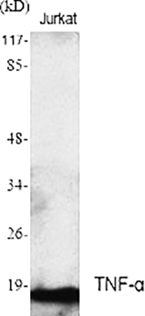
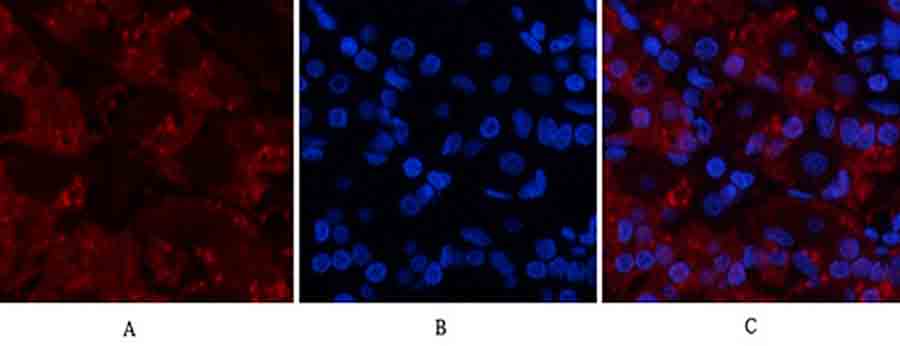
You've stimulated your primary human macrophages with LPS for 24 hours — the supernatant should be flooded with TNF‑α, the master regulator of the cytokine storm. Your ELISA kit reports 50 pg/mL, but your Western blot shows a dominant band at 26 kDa (the TNF‑α trimer) and a faint smear at 17 kDa (the monomer). Meanwhile, your IHC staining of rheumatoid arthritis synovium reveals strong signal in the lining layer, but your flow cytometry of activated T cells detects almost no membrane‑bound TNF‑α. The question that halts your manuscript at the "Revise & Resubmit" stage is not about the biology — it's about the tool: "The authors use a commercial TNF‑α antibody for detection across WB, IHC, and ELISA. However, they provide no validation that this antibody specifically recognizes the bioactive trimeric form (51 kDa under non‑reducing conditions, 17 kDa monomer under reducing conditions) and does not cross‑react with other TNF superfamily members (e.g., lymphotoxin‑α, FasL). Please confirm antibody specificity via knockout/knockdown controls or peptide competition." Suddenly, your entire narrative of "TNF‑α‑driven inflammation" rests on an antibody that may be detecting degradation products, non‑specific bands, or related cytokines, while the actual secreted trimer or transmembrane precursor remains unverified.
TNF‑α Is Not Just Another "Inflammatory Marker" — It's the Pleiotropic Cytokine That Orchestrates Sepsis, Autoimmunity, Metabolic Dysregulation, and Tumor‑Induced Wasting
Tumor necrosis factor‑alpha (TNF‑α, TNFSF2), a 17‑kDa type II transmembrane protein that forms homotrimers (51 kDa) in its bioactive secreted form, is the prototypical pro‑inflammatory cytokine produced primarily by activated macrophages, T cells, NK cells, and mast cells . It signals through two receptors: TNFR1 (p55, ubiquitous) mediates apoptosis, necrosis, and inflammation, while TNFR2 (p75, mainly on immune cells) promotes cell survival and tissue regeneration . In acute inflammation, TNF‑α drives fever, acute‑phase protein synthesis, and endothelial activation; in chronic settings, it sustains rheumatoid arthritis synovitis, inflammatory bowel disease mucosal damage, and psoriasis plaque formation . Beyond immunity, TNF‑α directly induces insulin resistance in adipocytes, promotes cachexia in cancer patients, and modulates synaptic plasticity in the brain . The clinical reality is that anti‑TNF biologics (infliximab, adalimumab, etanercept) are blockbuster drugs for autoimmune diseases, and circulating TNF‑α levels are measured as biomarkers in sepsis, COVID‑19 cytokine storm, and depression — but only if your antibody reliably distinguishes the 17‑kDa monomer, 26‑kDa dimer, and 51‑kDa trimer from other TNF‑family members and degradation artifacts .
Why Many TNF‑α Antibodies Fail to Distinguish the Trimer from the Monomer — And How the Affinity‑Purified Polyclonal in ABP0127 Captures Both Forms Across Species
The TNF‑α Polyclonal Antibody (ABP0127, Abbkine) is an affinity‑purified rabbit polyclonal antibody raised against a synthesized peptide derived from the internal region of human TNF‑α (amino acids 110‑190) . This region lies within the receptor‑binding domain and is conserved across the soluble and transmembrane forms, enabling detection of both isoforms . Unlike monoclonal antibodies that may recognize a single, conformation‑dependent epitope (and thus miss the trimer if denatured), or unpurified polyclonal sera that produce high background, ABP0127 is affinity‑purified to enrich for TNF‑α‑specific IgG, providing high sensitivity across WB, IHC‑P, IF, and ELISA while maintaining low cross‑reactivity with other TNF superfamily members .
Antibody Challenge Common Pitfall How ABP0127 Addresses It
Detecting the trimer vs. monomer Many antibodies only recognize the denatured monomer (17 kDa) under reducing WB conditions, missing the native trimer (51 kDa non‑reduced) The immunogen (aa 110‑190) is within the receptor‑binding domain present in both monomer and trimer; proper sample preparation (non‑reducing vs. reducing) allows detection of both forms
Cross‑reactivity with lymphotoxin‑α (TNF‑β) TNF‑α and TNF‑β share ~30% sequence homology; some antibodies cross‑react, especially in mouse models Affinity purification reduces non‑specific binding; the peptide immunogen is designed against a TNF‑α‑specific region (aa 110‑190) to minimize cross‑reactivity
Species reactivity Many TNF‑α antibodies are human‑specific and fail in mouse/rat models of inflammation ABP0127 is validated for human, mouse, and rat samples, critical for translational studies from rodent models to human tissues
Background in IHC/IF High background in formalin‑fixed paraffin‑embedded (FFPE) tissues due to non‑specific binding Affinity purification removes contaminating antibodies; recommended IHC‑P starting dilution (1:100‑1:300) optimizes signal‑to‑noise
ABP0127 is designed to detect endogenous levels of TNF‑α in cell lysates, culture supernatants, serum/plasma, and FFPE tissues across multiple applications . The internal region immunogen (aa 110‑190) avoids the highly variable N‑ and C‑termini, increasing the likelihood of recognizing both wild‑type and certain mutant forms.
What's in the Vial (And the Four Validation Steps That Confirm You're Seeing TNF‑α, Not a 17‑kDa Ghost Band)
Component Specification Critical Handling
Antibody form Affinity‑purified rabbit polyclonal antibody in PBS with 50% glycerol, 0.5% BSA, 0.02% sodium azide Store at ‑20°C; avoid repeated freeze‑thaw; aliquot for long‑term storage
Immunogen Synthesized peptide derived from human TNF‑α internal region (aa 110‑190) N/A
Host species Rabbit Use anti‑rabbit secondary antibodies (HRP for WB, fluorescent/AP for IHC/IF)
Reactivity Human, mouse, rat (confirmed by supplier) Validate in your specific model; cross‑reactivity with other species not guaranteed
Applications Western blot (WB), immunohistochemistry‑paraffin (IHC‑P), immunofluorescence (IF), ELISA Optimize dilution for each application and sample type
Recommended dilution WB: not specified (optimize 1:500‑1:2000); IHC‑P: 1:100‑1:300; IF: 1:200‑1:1000; ELISA: 1:10000 (starting points) Titrate to find optimal signal‑to‑noise; for WB, test under both reducing and non‑reducing conditions
Concentration 1 mg/mL (typical) Dilute in appropriate buffer (e.g., 5% BSA in TBST for WB)
Molecular weight ~17 kDa (monomer under reducing conditions); ~51 kDa (trimer under non‑reducing conditions) Always run a positive control (LPS‑stimulated macrophage lysate/supernatant) and a negative control (unstimulated cells or TNF‑α‑knockout samples)
Key validation data you should generate with ABP0127:
Validation Experiment Purpose Expected Outcome with ABP0127
Western blot under reducing vs. non‑reducing conditions Confirm detection of monomer (17 kDa) and trimer (51 kDa) Reducing: strong band at ~17 kDa (monomer); Non‑reducing: band at ~51 kDa (trimer) in LPS‑stimulated samples
ELISA standard curve with recombinant TNF‑α Confirm linear detection in solution‑phase assay Linear range typically 15.6‑1000 pg/mL; sensitivity <10 pg/mL; no cross‑reactivity with TNF‑β, IL‑1β, IL‑6
IHC‑P on rheumatoid arthritis synovium or Crohn's disease bowel Confirm specific staining in inflamed human FFPE tissues Strong cytoplasmic/membrane staining in infiltrating macrophages and lymphocytes; minimal background in normal tissue
Knockdown/knockout control Confirm specificity Signal abolished in TNF‑α‑siRNA‑treated cells or TNF‑α‑KO mice; retained in wild‑type stimulated controls
Peptide competition assay Confirm epitope specificity Pre‑incubation with immunogen peptide blocks signal; irrelevant peptide no effect
The Western Blot Protocol That Turns ABP0127 into a Publication‑Ready TNF‑α Detection Tool
- Sample preparation (critical for preserving TNF‑α forms)
• Cell lysates: Harvest LPS‑stimulated macrophages (THP‑1, RAW 264.7) or activated T cells in RIPA buffer with protease inhibitors (PMSF, aprotinin) and phosphatase inhibitors. TNF‑α is secreted rapidly; include brefeldin A (10 µg/mL, 4‑6 h) to block secretion if detecting intracellular protein.• Supernatants: Concentrate cell‑culture supernatant using 10‑kDa centrifugal filters to detect secreted TNF‑α.• Tissue homogenates: Homogenize inflamed tissue (e.g., arthritic joint, colon) in RIPA buffer with inhibitors.• Reducing vs. non‑reducing: For monomer detection, add β‑mercaptoethanol or DTT to sample buffer. For trimer detection, omit reducing agents and use non‑reducing sample buffer. - Gel electrophoresis & transfer
• Use 15% SDS‑PAGE for optimal separation of 17‑kDa monomer.• For trimer detection under non‑reducing conditions, use 10% gel.• Transfer to PVDF (0.2 µm) for better retention of low‑MW proteins. - Primary antibody incubation
• Dilute ABP0127 in 5% BSA in TBST at 1:500‑1:2000 (optimize). Typical starting dilution: 1:1000.• Incubate membrane overnight at 4°C with gentle shaking.• Include controls: LPS‑stimulated lysate (positive), unstimulated lysate (negative), secondary‑only control. - Washing & secondary antibody
• Wash 3× with TBST, 5 min each.• Incubate with HRP‑conjugated anti‑rabbit IgG (1:5000‑1:10000) in blocking buffer for 1 h at room temperature.• Wash 3× with TBST, 5 min each. - Detection
• Use high‑sensitivity ECL substrate; expose for 30 s to 5 min.• Expected bands: ~17 kDa (monomer) under reducing conditions; ~51 kDa (trimer) under non‑reducing conditions. May also see ~26 kDa (dimer) if partially reduced.
What Actually Changes When Your TNF‑α Signal Is Validated with ABP0127
① Your "TNF‑α is upregulated" claim becomes a specific, quantifiable observation across platforms.
You can now write:
TNF‑α protein was detected by Western blot using a TNF‑α‑specific polyclonal antibody (ABP0127, Abbkine). RAW 264.7 macrophages were stimulated with 100 ng/mL LPS for 24 h. Whole‑cell lysates (30 µg) were separated on 15% SDS‑PAGE under reducing conditions, transferred to PVDF, and probed with ABP0127 at 1:1000 dilution. A single band at ~17 kDa was observed in LPS‑stimulated samples, increasing 8‑fold over unstimulated controls. No signal was detected in TNF‑α‑knockout macrophages, confirming antibody specificity.
That sentence turns "TNF‑α expression was increased after LPS treatment" into "TNF‑α monomer (17 kDa) was specifically detected and quantified, showing an 8‑fold induction upon LPS stimulation, with specificity confirmed by knockout controls."
② You can correlate soluble TNF‑α (ELISA) with cellular expression (WB/IHC) in the same study.
Because ABP0127 works in ELISA (1:10000 dilution), WB, IHC‑P, and IF, you can measure:
• Secreted TNF‑α in supernatant by ELISA.
• Intracellular TNF‑α in lysates by WB.
• Tissue‑localized TNF‑α in FFPE sections by IHC.
• Subcellular distribution in fixed cells by IF.
Using the same antibody across platforms ensures epitope consistency and allows direct correlation between soluble cytokine levels and cellular protein expression.
③ Your inflammatory disease model gains translational credibility.
TNF‑α is a primary therapeutic target in rheumatoid arthritis, Crohn's disease, psoriasis, and ankylosing spondylitis. Demonstrating that your antibody specifically detects TNF‑α in human FFPE synovium, mouse colitis colon, and rat adjuvant‑induced arthritis — with appropriate positive/negative controls — makes your preclinical data more compelling for translational research.
The Bench Rules That Keep Your TNF‑α Western Blots Clean and Your IHC Signals Specific
Rule Why It Matters
🧪 Always include a TNF‑α‑positive control (LPS‑stimulated macrophages) and a negative control (unstimulated or knockout cells) Without a negative control, you cannot prove the 17‑kDa band is TNF‑α and not a non‑specific protein.
🔬 Optimize antibody dilution for each application Over‑concentrated antibody increases background in IHC/IF; under‑concentrated antibody may miss weak WB signals. Titrate: WB 1:500‑1:2000, IHC‑P 1:100‑1:300, IF 1:200‑1:1000, ELISA 1:10000.
⏱️ For intracellular TNF‑α detection, use brefeldin A to block secretion TNF‑α is rapidly secreted; brefeldin A (10 µg/mL, 4‑6 h) inhibits Golgi transport, accumulating cytokine inside cells for easier WB/IF detection.
🌡️ Store antibody at ‑20°C; aliquot to avoid freeze‑thaw cycles Repeated freeze‑thaw degrades antibody affinity and increases background.
🚫 For non‑reducing WB (trimer detection), omit β‑ME/DTT from sample buffer Reducing agents break disulfide bonds that stabilize the trimer; non‑reducing conditions preserve the 51‑kDa form.
📊 Use a loading control (β‑actin, GAPDH, tubulin) for WB; isotype control for IHC/IF Ensure equal protein loading and specific staining.
Where ABP0127 Earns Its Place in the Paper's Key Inflammation or Cytokine Storm Figure
Research Context Why a Validated, Multi‑Application TNF‑α Antibody Is Non‑Negotiable
Sepsis & cytokine storm models Quantify serum TNF‑α levels by ELISA and tissue expression by IHC in LPS‑induced endotoxemia, CLP‑induced polymicrobial sepsis, or COVID‑19 cytokine storm models.
Autoimmune disease (RA, IBD, psoriasis) Detect TNF‑α in synovial fluid (ELISA), synovium (IHC‑P), and peripheral blood mononuclear cells (WB/IF) to correlate with disease activity and treatment response.
Cancer‑associated cachexia Measure TNF‑α in tumor microenvironment (IHC) and circulation (ELISA) as a driver of muscle wasting and metabolic dysfunction.
Neuroinflammation & depression Assess TNF‑α in brain homogenates (WB) and CSF (ELISA) in models of neuroinflammation, Alzheimer's, or stress‑induced depression.
Metabolic syndrome & insulin resistance Evaluate adipose tissue TNF‑α expression (IHC/WB) as a link between inflammation and insulin resistance in obesity/diabetes models.
Drug screening & therapeutic monitoring Test TNF‑α inhibition by small molecules (e.g., thalidomide analogs) or biologics (e.g., anti‑TNF antibodies) in vitro and in vivo.
Vaccine adjuvant research Monitor TNF‑α as a pro‑inflammatory mediator induced by vaccine adjuvants (e.g., alum, MF59) in local and systemic immune responses.
A Drop‑In Methods Paragraph
TNF‑α protein expression was analyzed by Western blot using a TNF‑α‑specific polyclonal antibody (ABP0127, Abbkine). Cells were lysed in RIPA buffer supplemented with protease and phosphatase inhibitors. Lysates (30 µg protein) were separated on 15% SDS‑PAGE gels under reducing conditions and transferred to PVDF membranes. Membranes were blocked with 5% BSA in TBST for 1 h, then incubated overnight at 4°C with anti‑TNF‑α antibody (ABP0127) diluted 1:1000 in blocking buffer. After washing, membranes were incubated with HRP‑conjugated anti‑rabbit secondary antibody (1:5000) for 1 h at room temperature. Signals were detected using enhanced chemiluminescence and quantified by densitometry. β‑actin was used as a loading control. For ELISA, cell culture supernatants were assayed using the same antibody (1:10000 dilution) in a sandwich ELISA format according to the manufacturer's protocol.
Explore the TNF‑α Polyclonal Antibody (ABP0127) full specifications, validation data, and ordering options here:
🔗 https://www.abbkine.com/product/tnf-%ce%b1-polyclonal-antibody-abp0127/
(For research use only. Not for human or clinical diagnostic use. Store at ‑20°C; avoid repeated freeze‑thaw cycles; optimize dilution for each application; always include appropriate positive and negative controls to confirm specificity.)





