




Cell Migration Assay Kit (24-well, 8 μM) — Application Scenarios for Metastasis and Wound Healing Research
Cell Migration: A Critical Parameter in Cancer, Development, and Tissue Repair
Cell migration is a fundamental biological process underlying cancer metastasis, embryonic development, wound healing, and immune surveillance. Whether you're studying tumor cell invasiveness, evaluating the effects of chemotactic factors, or screening anti-metastatic compounds, reliable migration assays are essential.
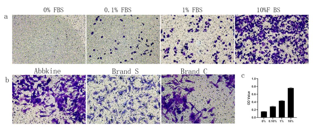
Abbkine's Cell Migration Assay Kit (24-well, 8 μM) (KTA5010) provides a straightforward, quantitative system for measuring cell migration through a porous membrane. Here's how researchers across different fields are putting it to work.
Cancer Research: Quantifying Metastatic Potential
Tumor cell migration is a prerequisite for metastasis. Cancer researchers use this kit to:
- Assess the migratory capacity of cancer cell lines (e.g., breast, lung, colon, melanoma)
- Evaluate the effects of candidate anti-metastatic compounds on cell motility
- Study the role of oncogenes and tumor suppressors in migration regulation
- Compare migration rates between primary and metastatic cell lines
The 8 μm pore size is ideal for most adherent cancer cell lines, allowing cells to actively migrate through the membrane in response to chemoattractants.
Immunology: Chemotaxis and Immune Cell Trafficking
Immune cell migration is central to inflammation, infection response, and autoimmunity. Immunologists leverage this kit to:
- Quantify chemotaxis of immune cells (macrophages, neutrophils, T cells) toward chemokines
- Study the effects of cytokines and inhibitors on immune cell motility
- Investigate leukocyte trafficking mechanisms
- Screen for immunomodulatory compounds that affect cell migration
Wound Healing and Regenerative Medicine
Cell migration is essential for tissue repair and regeneration. Researchers in wound healing use this kit to:
- Assess fibroblast and keratinocyte migration in wound healing models
- Evaluate the effects of growth factors (EGF, FGF, TGF-β) on cell motility
- Study stem cell homing and migration toward injured tissues
- Screen for pro-healing compounds that enhance cell migration
Developmental Biology: Understanding Morphogenetic Movements
Cell migration drives many morphogenetic processes during development. Developmental biologists apply this kit to:
- Study neural crest cell migration patterns
- Investigate gastrulation and other developmental movements
- Assess the effects of signaling pathways (WNT, TGF-β, FGF) on cell motility
- Characterize migration defects in developmental disorder models
Why Choose This Kit for Your Migration Research?
| Feature | Benefit |
|---|---|
| 24-well format | High-throughput compatibility; multiple conditions per experiment |
| 8 μM pore size | Optimal for most adherent cell types |
| Pre-coated membranes | Ready to use — no tedious coating steps |
| Quantitative readout | Reliable, reproducible migration data |
| Complete kit | All necessary components included |
| Proven design | Based on the classic Boyden chamber/transwell principle |
Key Experimental Considerations
- Cell type: Most adherent cell lines (cancer cells, fibroblasts, endothelial cells, immune cells)
- Chemoattractant: Serum, growth factors, chemokines, or conditioned media in the lower chamber
- Incubation time: Typically 4–24 hours depending on cell type and migration rate
- Readout: Stain and quantify migrated cells on the membrane underside
🔬 Which research field does your work belong to?
Cancer Research | Immunology | Wound Healing | Developmental Biology | Other
Share your application with us — we'd love to hear how you're using this kit in your studies!
📌 Reference Link: Cell Migration Assay Kit (24-well, 8 μM) — KTA5010





