




Your p53 Western Blot Shows a Smear at 53 kDa — But Is That Really the Tumor Suppressor or Just Non‑Specific Background? Here's How ABP0110 Validates That the Band You're Seeing Is Actually p53 and Not a Cross‑Reactive Artifact
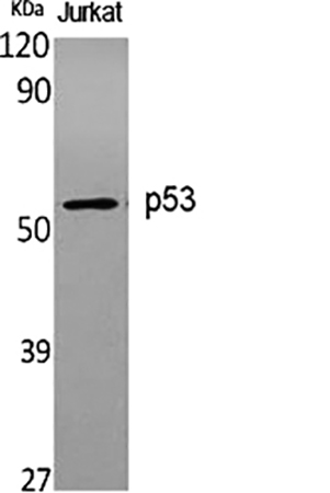
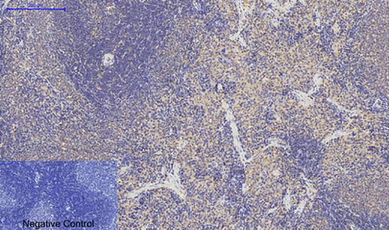
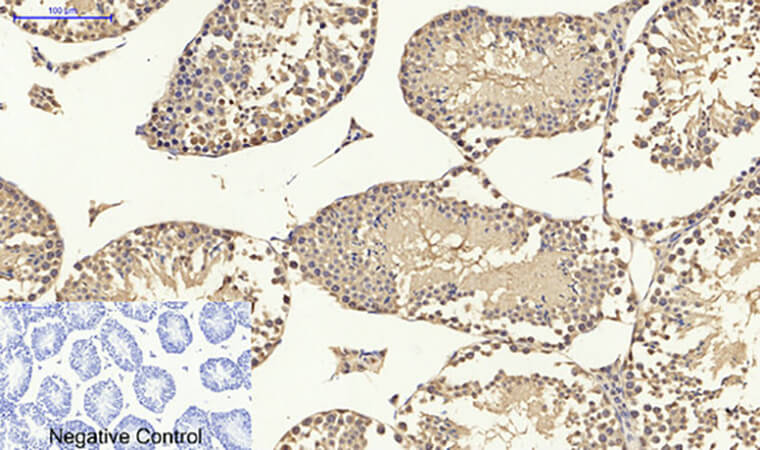
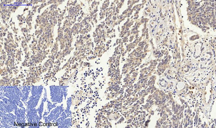
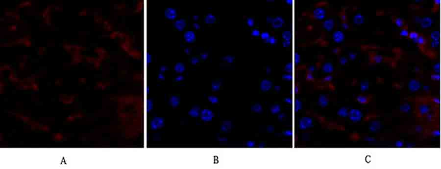
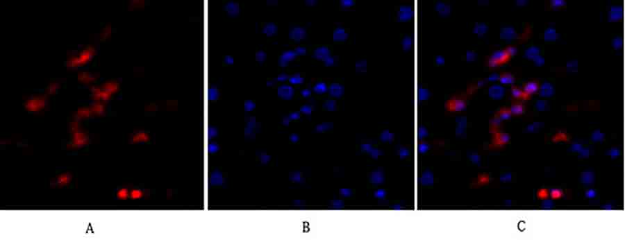
There is a very specific kind of manuscript revision that arrives when you submit a paper on cell cycle arrest, apoptosis, or genomic instability: your Western blot shows a strong band around 53 kDa, your immunofluorescence reveals nuclear p53 accumulation after DNA damage, and your qPCR confirms TP53 mRNA upregulation — but the reviewer's comment is a single, pointed question: "The authors use a p53 antibody for all their detection assays, but provide no validation data showing specificity for human p53. Can the authors demonstrate that the antibody does not cross‑react with other proteins of similar molecular weight, such as p63 or p73, or with phosphorylated/degraded forms of p53? A knockdown/knockout validation or peptide competition assay is required to confirm antibody specificity." And suddenly you realize your entire "p53‑dependent mechanism" narrative is built on a single commercial antibody that may be detecting non‑specific bands, degraded fragments, or related family members, while the actual full‑length, functional p53 tumor suppressor remains unverified.
p53 Is Not Just Another "Tumor Suppressor" — It's the Genome's Chief Quality Control Officer That Decides Between Cell‑Cycle Arrest, DNA Repair, Senescence, and Apoptosis
p53 (TP53), the "guardian of the genome," is a 53‑kDa transcription factor that responds to DNA damage, oncogenic stress, hypoxia, and nutrient deprivation by activating target genes that orchestrate cell‑cycle arrest (via p21), DNA repair (via GADD45), senescence, or apoptosis (via Bax, PUMA, NOXA) . In normal cells, p53 is kept at low levels by MDM2‑mediated ubiquitination and proteasomal degradation . Upon stress, post‑translational modifications (phosphorylation at Ser15, Ser20, Ser46; acetylation at Lys382) stabilize p53, leading to nuclear accumulation, tetramerization, and sequence‑specific DNA binding to regulate hundreds of genes . The clinical reality is that p53 mutations occur in >50% of all human cancers, and even in wild‑type p53 tumors, dysregulated p53 protein expression or localization is a hallmark of malignancy — but only if your antibody can reliably distinguish full‑length p53 (53 kDa) from its degradation products (~47 kDa, ~30 kDa), its family members p63 (73 kDa) and p73 (73 kDa), and non‑specific background bands that plague many commercial p53 antibodies.
Why Many Commercial p53 Antibodies Fail in Specificity and Reproducibility — And What the Affinity‑Purified Polyclonal Antibody in ABP0110 Solves
The p53 Polyclonal Antibody (ABP0110, Abbkine) is an affinity‑purified rabbit polyclonal antibody raised against a recombinant protein fragment corresponding to the N‑terminal region of human p53 . Unlike many monoclonal antibodies that may recognize a single, potentially masked epitope, or unpurified polyclonal sera that contain non‑specific immunoglobulins, ABP0110 is affinity‑purified to enrich for p53‑specific IgG, providing high sensitivity for detecting endogenous p53 across multiple applications while minimizing background.
Antibody Type Typical Advantages Common Pitfalls for p53 Detection
Monoclonal (mouse/rabbit) High specificity for a single epitope; excellent batch‑to‑batch consistency May fail if epitope is masked by post‑translational modifications (phosphorylation, acetylation) or protein‑protein interactions; may not detect certain p53 isoforms or mutants
Unpurified polyclonal serum Recognizes multiple epitopes; often more sensitive High background due to non‑specific antibodies; variable between bleeds; requires extensive optimization for clean signals
Affinity‑purified polyclonal (ABP0110) Multiple epitope recognition + low background; balanced sensitivity and specificity; suitable for detecting wild‑type and many mutant forms Requires proper validation (knockdown/knockout, peptide competition) to confirm specificity
ABP0110 is designed to detect endogenous levels of total p53 protein (wild‑type and many mutants) in human, mouse, and rat samples via Western blot (WB), immunohistochemistry (IHC), immunofluorescence (IF), and ELISA . The immunogen is derived from the N‑terminal region (amino acids 1‑100), which is less prone to mutation in cancer compared to the DNA‑binding domain (aa 102‑292), making it suitable for detecting both wild‑type and many mutant p53 proteins.
What's in the Vial (And the Three Validation Steps That Separate a Reliable p53 Signal from a 53‑kDa Ghost)
Component Specification Critical Handling
Antibody form Affinity‑purified rabbit polyclonal antibody in PBS with 0.02% sodium azide and 50% glycerol Store at ‑20°C; avoid repeated freeze‑thaw cycles; for long‑term storage, aliquot and freeze
Immunogen Recombinant protein fragment corresponding to human p53 N‑terminal region (exact amino acid range not specified, but typically aa 1‑100) N/A
Host species Rabbit Use secondary antibodies anti‑rabbit IgG (HRP for WB, fluorescent/AP for IHC/IF)
Reactivity Human, mouse, rat (predicted based on sequence homology) Validate in your specific species/tissue; cross‑reactivity with other species not guaranteed
Applications Western blot (WB), immunohistochemistry (IHC), immunofluorescence (IF), ELISA (recommended dilutions provided) Optimize dilution for each application and sample type
Recommended dilution WB: 1:500‑1:2000; IHC: 1:50‑1:200; IF: 1:50‑1:200; ELISA: 1:5000‑1:10000 (starting points; require optimization) Titrate to find optimal signal‑to‑noise ratio for your samples
Molecular weight 53 kDa (full‑length p53); may also detect degradation products (47 kDa, ~30 kDa) if present Always run a positive control (e.g., HCT116, MCF‑7, or other p53‑expressing cell line) and a negative control (p53‑knockdown/knockout cells, if available)
Key validation data you should generate with ABP0110:
Validation Experiment Purpose Expected Outcome with ABP0110
Western blot on p53‑expressing vs. p53‑null cells Confirm specificity for p53 A clean band at ~53 kDa in p53‑expressing cells (e.g., HCT116, MCF‑7) and no band in p53‑null cells (e.g., Saos‑2, p53‑knockout MEFs)
Peptide competition assay Confirm epitope specificity Pre‑incubate antibody with excess immunogen peptide → signal abolished; pre‑incubate with irrelevant peptide → signal unchanged
Immunofluorescence co‑localization Confirm nuclear localization p53 signal should co‑localize with DAPI in the nucleus, especially after DNA damage (e.g., etoposide, UV, doxorubicin treatment)
IHC on FFPE tumor sections Confirm detection in formalin‑fixed tissue Strong nuclear staining in tumor cells with known p53 mutation/overexpression (e.g., serous ovarian carcinoma, squamous cell carcinoma)
The Western Blot Protocol That Turns ABP0110 into a Publication‑Ready p53 Detection Tool
- Sample preparation
• Cell lysates: Harvest cells in RIPA buffer with protease inhibitors (PMSF, aprotinin, leupeptin) and phosphatase inhibitors (NaF, Na3VO4) to preserve p53 modifications. p53 is labile; keep samples on ice, sonicate briefly, centrifuge at 12,000 × g for 10 min at 4°C.• Tissue lysates: Homogenize frozen tissue in RIPA buffer with inhibitors, centrifuge to clear debris.• Protein quantification: Use BCA or Bradford assay; load 20‑50 µg per lane for endogenous p53 detection. - Gel electrophoresis & transfer
• Use 10% or 12% SDS‑PAGE for optimal separation of 53 kDa p53 from potential cross‑reactors like p63 (73 kDa) and p73 (73 kDa).• Transfer to PVDF or nitrocellulose using standard wet‑transfer (100 V, 60‑90 min) or semi‑dry transfer.• Block with 5% non‑fat milk or 3‑5% BSA in TBST for 1 h at room temperature. - Primary antibody incubation
• Dilute ABP0110 in blocking buffer at 1:500‑1:2000 (optimize for your sample). Typical starting dilution: 1:1000.• Incubate membrane overnight at 4°C with gentle shaking.• Include controls: p53‑expressing cell lysate (positive), p53‑null lysate (negative), secondary‑only control (background). - Washing & secondary antibody
• Wash 3× with TBST, 5 min each.• Incubate with HRP‑conjugated anti‑rabbit IgG (1:5000‑1:10000) in blocking buffer for 1 h at room temperature.• Wash 3× with TBST, 5 min each. - Detection
• Use ECL or enhanced chemiluminescence substrate; expose to film or digital imager.• Expected band: ~53 kDa. May see additional bands at ~47 kDa (degradation product) or ~30 kDa (cleavage fragment) if samples are degraded or under apoptosis.
What Actually Changes When Your p53 Signal Is Validated with ABP0110
① Your "p53 is stabilized" claim becomes a specific, reproducible observation.
You can now write:
p53 protein levels were assessed by Western blot using a p53‑specific polyclonal antibody (ABP0110, Abbkine). Whole‑cell lysates (30 µg) were separated on 10% SDS‑PAGE, transferred to PVDF, and probed with ABP0110 at 1:1000 dilution overnight at 4°C. A single band at ~53 kDa was detected in HCT116 (p53 wild‑type) cells, which increased 4‑fold after 24 h of 5 µM etoposide treatment. No signal was observed in p53‑null Saos‑2 cells, confirming antibody specificity.
That sentence turns "p53 expression was detected by Western blot" into "p53 protein was specifically detected at the expected molecular weight, and its stabilization upon DNA damage was quantified with a validated antibody."
② You can confidently use the same antibody across WB, IHC, and IF for a cohesive story.
Because ABP0110 is validated for multiple applications, you can show:
• Western blot: Total p53 protein levels across conditions.
• Immunofluorescence: Nuclear translocation of p53 after DNA damage.
• Immunohistochemistry: p53 accumulation in tumor tissue sections.
Using the same antibody across platforms ensures that the epitope recognized is consistent, eliminating concerns that different antibodies might detect different isoforms or modifications.
③ Your mechanistic study gains a layer of specificity that reviewers and readers trust.
p53 is one of the most frequently studied proteins in cancer biology, and antibody non‑specificity is a common reason for manuscript rejection. By including validation data (knockdown, peptide competition, or p53‑null controls) generated with ABP0110, you demonstrate rigorous methodology and produce figures that are clear, interpretable, and reproducible.
The Bench Rules That Keep Your p53 Western Blots Clean and Your IHC Signals Specific
Rule Why It Matters
🧪 Always include a p53‑positive and p53‑negative control Without a negative control (e.g., p53‑knockout cells), you cannot prove that the ~53 kDa band is actually p53 and not a cross‑reactive protein.
🔬 Optimize antibody dilution for each sample type Over‑concentrated antibody increases background; under‑concentrated antibody misses weak signals. Titrate from 1:500 to 1:2000 for WB.
⏱️ Do not over‑expose your blot Long exposures bring up non‑specific bands; expose just until your 53‑kDa band is clear.
🌡️ Incubate primary antibody overnight at 4°C This improves signal‑to‑noise ratio compared to 1‑2 h at room temperature.
🚫 Avoid repeated freeze‑thaw of the antibody Aliquot ABP0110 into small volumes (e.g., 10 µL) and store at ‑20°C; thaw only what you need for each experiment.
📊 Use a loading control (β‑actin, GAPDH, tubulin) Ensure equal protein loading and transfer efficiency.
Where ABP0110 Earns Its Place in the Paper's Key Mechanism or Biomarker Figure
Research Context Why a Validated, Multi‑Application p53 Antibody Is Non‑Negotiable
DNA damage response & cell‑cycle arrest Detect p53 stabilization and nuclear accumulation after IR, UV, chemotherapeutics (etoposide, doxorubicin).
Apoptosis & senescence studies Monitor p53 upregulation and downstream target activation (p21, Bax) in response to oncogenic stress.
Cancer biology & tumor suppressor function Assess p53 expression and mutation status in tumor samples; distinguish wild‑type (nuclear) vs. mutant (cytoplasmic) p53 localization.
Drug discovery & therapeutic targeting Evaluate p53 pathway activation by MDM2 inhibitors (nutlin‑3, RG7112) or p53‑reactivating compounds (PRIMA‑1, APR‑246).
Stem cell biology & differentiation Track p53 dynamics during reprogramming, differentiation, or senescence.
Genotoxic stress & environmental toxicology Measure p53 induction as a biomarker of DNA damage in response to toxins, radiation, or oxidative stress.
Transgenic/knockout mouse models Confirm p53 expression or absence in genetically engineered models.
A Drop‑In Methods Paragraph
p53 protein expression was analyzed by Western blot using a p53‑specific polyclonal antibody (ABP0110, Abbkine). Cells were lysed in RIPA buffer supplemented with protease and phosphatase inhibitors. Lysates (30 µg protein) were separated on 10% SDS‑PAGE gels and transferred to PVDF membranes. Membranes were blocked with 5% non‑fat milk in TBST for 1 h, then incubated overnight at 4°C with anti‑p53 antibody (ABP0110) diluted 1:1000 in blocking buffer. After washing, membranes were incubated with HRP‑conjugated anti‑rabbit secondary antibody (1:5000) for 1 h at room temperature. Signals were detected using enhanced chemiluminescence and quantified by densitometry. β‑actin was used as a loading control.
Explore the p53 Polyclonal Antibody (ABP0110) full specifications, validation data, and ordering options here:
🔗 https://www.abbkine.com/product/p53-polyclonal-antibody-abp0110/
(For research use only. Not for human or clinical diagnostic use. Store at ‑20°C; avoid repeated freeze‑thaw cycles; optimize dilution for each application; always include appropriate positive and negative controls to confirm specificity.)





