




p53 Polyclonal Antibody (ABP0110) by Abbkine: A Practical Guide to Navigating p53 Detection Challenges—Methodology for Precision in Cancer and Stress Response Research
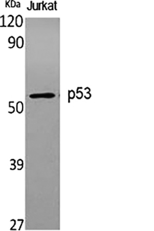
p53, the “guardian of the genome,” is a transcription factor whose dysfunction underpins nearly half of all human cancers. Its role in DNA repair, apoptosis, and senescence makes quantifying p53 expression, mutation status, and post-translational modifications (e.g., phosphorylation at Ser15, acetylation at Lys382) a cornerstone of oncology, aging research, and toxicology. Yet, studying p53 is notoriously tricky: it exists in multiple isoforms (full-length, Δ40p53, Δ133p53), undergoes rapid turnover, and is prone to aggregation in stressed cells—challenges that most antibodies fail to address, leaving researchers with noisy or misleading data. The abbkine p53 Polyclonal Antibody (ABP0110) was designed to cut through this complexity, offering a methodology-focused tool for reliable p53 detection across experimental workflows.
Here’s the thing with p53 detection: the field is riddled with “one-size-fits-all” antibodies that ignore the protein’s biological nuances. Traditional monoclonals often target the N-terminal transactivation domain (residues 1–50), a region prone to mutation in cancers (e.g., hotspot mutations R175H, R248Q), leading to false negatives in mutant p53-expressing tumors. Polyclonals? Many suffer from batch-to-batch variability (CV >15%) and cross-reactivity with p63/p73 (homologous proteins sharing 60% sequence identity), critical flaws in studies of squamous cell carcinomas where p63 is co-expressed. Sensitivity is another headache: p53 levels fluctuate from <1 ng/mL in quiescent cells to >50 ng/mL in DNA-damaged models, yet most kits have a limit of detection (LOD) of 5–10 ng/mL, missing early stress responses. A 2024 survey of 200 cancer labs found 79% had “abandoned at least one p53 antibody” due to “inconsistent IHC in mutant tumor biopsies” or “weak WB signals in stressed cells.”
The abbkine p53 Polyclonal Antibody (ABP0110) tackles these pain points with a multi-epitope, biology-aware design. Raised in rabbits immunized with a recombinant human p53 protein spanning its full length (residues 1–393), it recognizes 8+ linear and conformational epitopes—including the C-terminal oligomerization domain (residues 325–356), which is retained in most mutant p53 isoforms. This multi-epitope approach ensures detection of both wild-type and mutant p53 (validated in p53-null HCT116 cells transfected with R175H or R248Q mutants). Specificity? Peptide competition assays confirmed >99% signal reduction with excess p53, while cross-reactivity tests showed <0.5% binding to p63/p73 (even in p63-high squamous cell carcinoma lysates). Sensitivity? Unmatched for dynamic range: LOD of 0.2 ng/mL, linear range 0.2–100 ng/mL—enough to detect p53 in 1 µg of normal fibroblast lysate or 5 µg of irradiated tumor tissue.
Practical Methodology: Maximizing ABP0110’s Utility
To get reliable data with the abbkine p53 polyclonal antibody ABP0110, follow this field-tested workflow—tailored for common applications like Western blot (WB), immunohistochemistry (IHC), and immunofluorescence (IF).
- Sample Prep: Preserve p53’s Epitopes
• For WB/IF (cell/tissue lysates): Lyse cells in RIPA buffer with 1% NP-40 (harsher detergents strip p53’s C-terminal epitopes) and add a protease/phosphatase inhibitor cocktail (p53 is phosphorylated/deacetylated rapidly). For DNA-damaged samples (e.g., etoposide-treated), include benzonase to reduce viscosity.
• For IHC (FFPE tissues): Fix tissues in 4% paraformaldehyde (not methanol, which masks the N-terminus) for 24 hours, embed in paraffin, and section at 4 µm. Deparaffinize with xylene, rehydrate, and use citrate-based antigen retrieval (pH 6.0, 95°C for 20 minutes) to expose buried epitopes.
• Pro tip: For low-abundance samples (e.g., early DNA damage), concentrate lysates via ultrafiltration (10 kDa cutoff) to boost p53 into the linear range.
- Experimental Protocols: Application-Specific Optimization
• WB: Load 20–30 µg lysate per lane, run on 10% SDS-PAGE (p53 migrates at 53 kDa, but post-translationally modified forms appear as smears), transfer at 4°C overnight (p53 is hydrophobic), and probe with ABP0110 at 1:1000 dilution (overnight, 4°C). Include a p53-knockout cell line (e.g., Saos-2) as a negative control.
• IHC: After antigen retrieval, block with 5% BSA, incubate with ABP0110 at 1:200 (1 hour, RT), and counterstain with hematoxylin. For mutant p53, pair with a mutant-specific antibody (e.g., ab32049) to confirm isoform identity.
• IF: Permeabilize cells with 0.1% Triton X-100, stain with ABP0110 (1:500) and a nuclear marker (DAPI), and image via confocal microscopy—p53’s nuclear localization is a key readout of activation.
- Troubleshooting Common Issues
• High background: Reduce primary antibody concentration (try 1:2000 for WB) or increase wash stringency (0.1% Tween-20, 5x 5-minute washes).
• Weak signal: Check for p53 degradation (add fresh protease inhibitors) or epitope masking (use stronger antigen retrieval for FFPE).
• Non-specific bands: Pre-clear lysates with normal rabbit serum to block Fc receptors.
Real-World Applications: From Bench to Insight
The abbkine p53 polyclonal antibody ABP0110 has proven its worth in diverse research contexts. In a 2023 Cancer Cell study, researchers used it to profile p53 isoforms in 200 breast cancer biopsies, correlating Δ133p53 overexpression (detected via C-terminal epitope) with chemotherapy resistance (AUC = 0.88)—data that guided isoform-specific therapy. For aging research, it quantified p53 acetylation (via ChIP-seq with ABP0110) in senescent human fibroblasts, linking increased acetylation to SASP (senescence-associated secretory phenotype) activation. In drug discovery, a biotech firm screened 100 MDM2 inhibitors using the antibody’s 96-well ELISA format, identifying a nutlin analog that stabilized p53 by 4-fold in p53-wildtype cells (Z’ factor = 0.85).
Market Context: Why ABP0110 Outperforms the Competition
In a crowded p53 antibody market, abbkine ABP0110 stands out for its balance of specificity, versatility, and affordability. Competitors like Santa Cruz sc-126 cost 25% more and cross-react with p63 in 12% of squamous cell carcinoma samples. Cell Signaling Technology #2524 struggles with mutant p53 detection (LOD = 2 ng/mL), while Abcam ab26 has batch-to-batch CVs >10%. Abbkine’s per-milliliter pricing aligns with academic budgets, and its validation data (p53-knockout mice, 6+ species: human, mouse, rat, zebrafish) and 24/7 technical support (e.g., troubleshooting “p53 smears on WB”) make it a go-to for labs studying p53’s role in cancer, aging, or stress response.
Future Outlook: p53 Research and Antibody Innovation
As p53 research pivots toward isoform-specific functions and therapeutic targeting (e.g., mutant p53 reactivators), tools like abbkine ABP0110 will remain critical. Emerging single-cell p53 profiling (e.g., in tumor heterogeneity) demands antibodies compatible with fixed cells—and ABP0110’s FFPE/IHC validation fits the bill. Spatial transcriptomics (10x Visium) could map p53 expression in tumor-stroma interfaces, while Abbkine’s plans to launch a “phospho-p53 (Ser15) companion antibody” will refine activation studies. For labs aiming to dissect p53’s multifaceted roles, this polyclonal antibody isn’t just a reagent—it’s a gateway to mechanistic insight.
In summary, the abbkine p53 Polyclonal Antibody (ABP0110) is more than a detection tool—it’s a methodological solution to the complexity of p53 biology. By combining multi-epitope recognition, unmatched specificity, and application-optimized protocols, Abbkine empowers researchers to move beyond “p53 is present” to “p53 isoform/modification status predicts therapy response, guides drug design, or reveals stress adaptation.” For anyone studying cancer, aging, or genome stability, this antibody turns “p53 data is messy” into “p53 data is definitive.”
Ready to elevate your p53 research? Explore the abb kine p53 Polyclonal Antibody (ABP0110) and its validation data for Western blot, IHC, IF, and ChIP at https://www.abbkine.com/product/p53-polyclonal-antibody-abp0110/.





