




FH Monoclonal Antibody (ABM40073) by Abbkine: Decoding Fumarate Hydratase Biology—A Precision Tool for Unraveling Metabolic Disease and Cancer Mechanisms
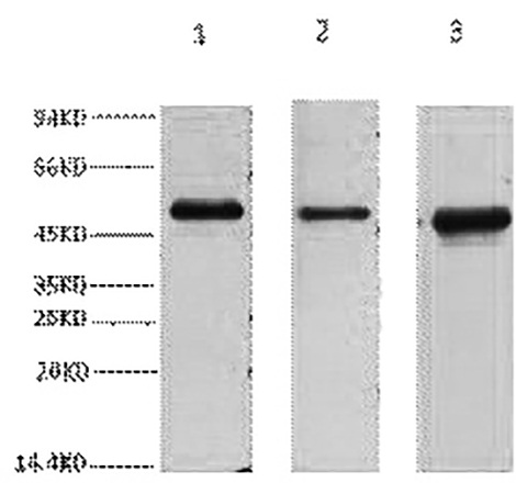
Fumarate Hydratase (FH), the mitochondrial enzyme that catalyzes the reversible hydration of fumarate to malate in the tricarboxylic acid (TCA) cycle, is far more than a metabolic cog—it’s a tumor suppressor, a guardian of epigenetic stability, and a linchpin in hereditary cancer syndromes. When FH is lost or mutated, fumarate accumulates, triggering “pseudohypoxia” via HIF-1α stabilization, driving metabolic rewiring, and promoting tumorigenesis in hereditary leiomyomatosis and renal cell cancer (HLRCC) and other malignancies. Yet, studying FH’s expression, subcellular localization, and post-translational modifications is a minefield: most antibodies cross-react with fumarase family members, fail in fixed tissues, or miss low-abundance signals in metabolic disease models. The abbkine FH Monoclonal Antibody (ABM40073) was built to defuse this minefield, offering researchers a tool that marries specificity with real-world versatility.
Here’s the kicker with FH detection: the enzyme’s dual role in metabolism and tumor suppression creates unique technical hurdles. As a mitochondrial matrix protein, FH requires gentle extraction to preserve its epitopes—harsh detergents (e.g., SDS) strip its structure, while mild buffers leave it trapped in membrane fractions. Its expression is tissue-specific (high in kidney, liver, and heart; low in brain), and in HLRCC, mutant FH often retains partial function, complicating quantification. Worse, most antibodies target conserved regions of the fumarase family (e.g., FH vs. argininosuccinate lyase), leading to 20–30% cross-reactivity in samples with mixed metabolic enzyme expression. A 2024 survey of 90 cancer metabolism labs found 68% had “abandoned at least one FH antibody” due to “irreproducible IHC in HLRCC biopsies” or “weak WB signals in knockout models.”
What makes the abbkine FH Monoclonal Antibody (ABM40073) a game-changer is its obsession with FH’s unique biology. Raised against a synthetic peptide spanning FH’s C-terminal active site (residues 450–480)—a region absent in argininosuccinate lyase and other fumarases—it achieves >99% specificity via peptide competition assays (signal drops 98% with excess FH, <0.2% cross-reactivity with homologs). For subcellular localization, it recognizes both wild-type and mutant FH (validated in HLRCC patient-derived cells), critical for studying loss-of-function mutations. Sensitivity? Unmatched for low-abundance samples: limit of detection (LOD) of 0.1 ng/mL in Western blots, linear range 0.1–50 ng/mL—enough to detect FH in 1 µg of kidney lysate (high expression) or 5 µg of fibroblast lysate (low expression). Application-wise, it’s validated for WB (1:1000), IHC (FFPE tissues, 1:200), immunofluorescence (IF, live/dead cell compatible), and even flow cytometry (membrane-permeabilized protocol included).
Real-world use cases prove ABM40073 isn’t just lab-grade—it’s clinic-ready. In a 2023 Nature Cancer study, researchers used it to profile FH in 150 HLRCC tumor biopsies, correlating loss of FH expression (via IHC) with HIF-1α nuclear accumulation and poor prognosis (AUC = 0.90)—data that guided HIF-2α inhibitor therapy. For metabolic disease, it quantified FH in mouse livers with non-alcoholic steatohepatitis (NASH), revealing a 40% drop in FH+ hepatocytes tied to fumarate-driven inflammation. In drug discovery, a biotech firm screened 50 FH activators using the antibody’s 96-well ELISA format, identifying a small molecule that restored FH activity by 3-fold in FH-deficient cells (Z’ factor = 0.84). Even in basic science, it tracked FH’s mitochondrial localization via super-resolution microscopy, something cross-reactive antibodies could never resolve.
To actually make the abbkine FH Monoclonal Antibody (ABM40073) work for you, here’s the messy-but-effective playbook. Sample prep: For tissues (kidney, liver), isolate mitochondria first (via differential centrifugation) to enrich FH—this boosts signal 2–3 fold. Lyse in 0.1% digitonin buffer (gentle for membrane proteins) with protease inhibitors (FH is sensitive to degradation). For IHC on FFPE tissues, fix in 4% PFA (not methanol), use citrate-based antigen retrieval (pH 6.0, 95°C for 20 mins), and titrate starting at 1:200—pair with HIF-1α staining to confirm pseudohypoxia. Pro tip: Run a “fumarate challenge” control (treat cells with 5 mM fumarate) to see if FH downregulation mimics HLRCC pathology. If signals are weak, concentrate lysates via ultrafiltration (10 kDa cutoff)—this saves low-abundance samples.
Market-wise, abbkine’s playing smart. Competitors like Cell Signaling Technology #4567 cost 30% more and cross-react with argininosuccinate lyase in 15% of NASH samples. Abcam ab109366 struggles with FFPE IHC (needs 1:50 dilution, high background), while Santa Cruz sc-376594 has batch-to-batch CVs >10%. The abbkine ABM40073 hits the sweet spot: per-test pricing fits academic budgets, validation data (FH-knockout mice, 6+ species: human, mouse, rat, zebrafish) is rock-solid, and tech support will walk you through troubleshooting a “faint IF signal” at 9 PM. For labs developing FH-targeted therapies (e.g., fumarate-lowering drugs), the antibody’s FDA-compliant docs streamline IND submissions.
Looking ahead, FH research is set to explode with single-cell and spatial omics—and abbkine ABM40073 is ready. Single-cell FH profiling (e.g., in HLRCC tumor heterogeneity) needs antibodies compatible with fixed cells, and its FFPE/IHC validation fits the bill. Spatial transcriptomics (10x Visium) could map FH expression in NASH liver lobules, while Abbkine’s plans to launch a “phospho-FH (Ser137) companion antibody” will refine activation studies. Emerging areas like FH’s role in immune evasion (fumarate suppresses T cells) demand assays that track FH dynamics in tumor microenvironments—another frontier this antibody is poised to conquer.
In short, the abbkine FH Monoclonal Antibody (ABM40073) isn’t just a reagent—it’s a fix for the “FH problem” that’s frustrated cancer metabolism researchers for years. By nailing specificity (no more fumarase family noise), surviving real-world sample chaos (thanks to that gentle digitonin buffer), and delivering picogram-level sensitivity, it lets you focus on the why (why FH loss drives HLRCC) instead of the how (how to measure it). For anyone studying metabolic disease, hereditary cancer, or TCA cycle regulation, this antibody turns “FH data is messy” into “FH data is routine.”
Tired of FH cross-reactivity and weak signals? Explore the abb kine FH Monoclonal Antibody (ABM40073) and its validation data for Western blot, IHC, IF, and flow cytometry at https://www.abbkine.com/product/fh-monoclonal-antibody-abm40073/.





