


More Than Just a Water Pipe: Why AQP3's Hidden Talent for Glycerol & H₂O₂ Makes It the Skin, Kidney, and Tumor Metabolic Switch You Keep Measuring Wrong — And How KTE62203 Finally Puts a Number on the Basolateral Channel
Every textbook intro to aquaporins draws you the same cartoon: a dumbbell-shaped tetramer with a narrow pore that lets water zip through a lipid bilayer at ~10⁹ molecules/sec, and then moves on to AQP1 in RBCs and AQP2 in vasopressin-regulated collecting ducts. But AQP3 (Aquaporin 3, aliases AQP-3, GIL/gill blood group, UniProt: Q92482, Gene ID: 360, Chr 9p13.3, ~292 aa / computed ~31.5 kDa) refuses to stay in that tidy "water-only" box. It belongs to the aquaglyceroporin subfamily — meaning its pore is just wide enough, and its selectivity filter just permissive enough, to cotransport glycerol and urea alongside water, and — the twist that rewrote the field — H₂O₂ (hydrogen peroxide) at rates that make AQP3 a redox signaling conduit, not just a plumbing fitting. In the skin, that means epidermal glycerol import → phospholipid biosynthesis → barrier repair. In the kidney, it means basolateral glycerol/urea recycling that supports concentrating capacity. In cancer, it means AQP3-dependent glycerol metabolism → Warburg-compatible carbon shunt + H₂O₂-mediated proliferation signaling. The Human Aquaporin 3 (AQP3) ELISA Kit (KTE62203) from Abbkine is the reagent that lets you measure this multitasking membrane channel as a calibrated sandwich-ELISA variable (ng/mL) — so your skin-hydration, renal-concentrating, or tumor-metabolism story rests on a number, not a "band at ~27–40 kDa that might be glycosylated or might be AQP4."
AQP3 in One Paragraph: The ~31-kDa Tetramer That Carries Water, Glycerol, and Oxidant Signals Across the Basolateral Membrane
The AQP3 monomer folds as a six-transmembrane-span (TM1–TM6) homotetramer, each subunit contributing its own pore lined by the conserved NPA (Asn-Pro-Ala) boxes that seal the water wire. But unlike AQP1/AQP2 — which have an aromatic/Arg constriction too tight for anything larger than water — AQP3's selectivity filter tolerates neutral solutes up to ~92 Da, opening the permeability hierarchy:
Substrate Relative Permeability (AQP3 vs. AQP1) Biological Payoff
H₂O ★★★ (high) Basolateral water flux in collecting duct, skin hydration
Glycerol ★★☆ (detectable) Keratinocyte phospholipid synthesis, renal medullary glycerol shuttle
Urea ★☆☆ (low) Supports inner-medullary urea recycling (antidiuresis)
H₂O₂ ★☆☆ (functionally critical) Redox signaling: AQP3 imports extracellular H₂O₂ → modulates EGFR/Mapk, PP2A oxidation, and proliferative tone in keratinocytes & fibroblasts
The translational localizations are equally specific: basolateral membrane of principal cells in the CCD (collecting duct), basal layer of stratified squamous epithelia (epidermal keratinocytes), urothelium, conjunctival epithelium, and respiratory epithelium — always the side facing the interstitium, never the apical urinary/luminal face (that's AQP2's job).
Why a Sandwich ELISA for a ~27–40 kDa Multi-Pass Membrane Protein — And Why "Just WB the 27K Band" Leaves Money on the Table
AQP3 is integral membrane (6TM) and runs as a doublet on reducing SDS-PAGE:
• ~27–31 kDa (core non-glycosylated monomer, sometimes called the "mature" form depending on tissue)
• ~37–42 kDa (N-linked glycosylated form — Asn¹³⁴ → N-glycan — more prominent in skin and some epithelial lines; deglycosylates with PNGaseF down to ~27 kDa)
That doublet is the first trap: without a two-epitope sandwich (pre-coated capture + biotin detection, different face of the extramembrane loop), you're always one bad transfer or one over-loaded lane away from misreading the ratio. Add the second trap — AQP3 levels are regulated transcriptionally (PPARγ/δ, HDAC3, p53, osmotic stress) AND by trafficking (vasopressin-independent translocation to/from the membrane) — and "band vs. actin" becomes an incomplete claim about a trafficking protein whose location matters as much as its mass.
The KTE62203 kit uses the proven two-site architecture:
- Microplate pre-coated with capture anti-AQP3 (raised against an extracellular / accessible loop epitope that survives both glycosylated and non-glycosylated forms).
- Standards (recombinant human AQP3) + samples — serum/plasma (exploratory; most AQP3 is membrane-integral), tissue homogenates, cell lysates, cell culture supernatants/lysates, other biological fluids — added → AQP3 (solubilized epitope pool) binds.
- Wash → biotinylated anti-AQP3 detection (different epitope) → Streptavidin–HRP.
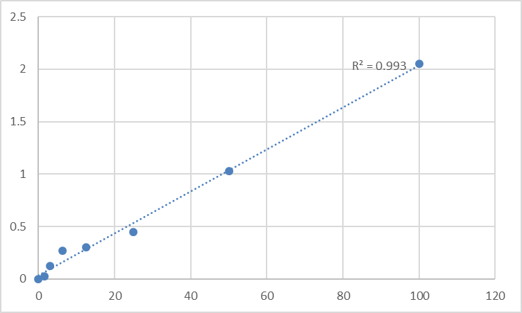
- TMB → stop → 450 nm → interpolate AQP3 concentration from the standard curve.
Typical performance envelope confirmed across distributor/technical references in this class:
Parameter KTE62203-class Specification
Target Human AQP3 / Aquaglyceroporin-3 (UniProt Q92482, Gene 360)
Format 96-well sandwich ELISA, pre-coated capture
Detection Biotin-Ab → SA-HRP → TMB, 450 nm
Dynamic Range 0.312 – 20 ng/mL
Sensitivity / LOD ~0.126–0.188 ng/mL
Intra-Assay CV < 8%
Inter-Assay CV < 10–12%
Specificity No significant cross-reactivity with AQP1, AQP2, AQP4, AQP5 or related MIP family at physiological levels
Samples Serum/plasma (exploratory), tissue homogenates, cell lysates, culture supernatants
Assay time ~3–5 hours
(Confirm exact dilutions and lot-specific recovery on the shipped Abbkine datasheet/CoA for KTE62203.)
Where AQP3 Quantification Actually Advances the Paper
- Skin Hydration, Barrier Repair & the "Cosmeceutical" Evidence Gap
This is where AQP3 earned its human-interest fame. AQP3-knockout mice have dry, flaky skin, reduced epidermal glycerol, impaired barrier-recovery after tape-strip, and — critically — blunted proliferative response to tumor promoters (TPA), because the glycerol–PLD2–PG (phosphatidylglycerol) axis that drives keratinocyte differentiation and lipid synthesis depends on imported glycerol. Conversely, epidermal AQP3 overexpression speeds barrier recovery.
The actionable readout for any skin-biology or topical-formulation lab: quantify AQP3 in keratinocyte/epidermal lysates (ng/mg, BCA) alongside TEWL (transepidermal water loss), stratum corneum hydration (Corneometer), glycerol content (LC-MS or enzymatic), and differentiation markers (loricrin, filaggrin, involucrin). That's the triad that moves the claim from "skin looks better" to "the glycerol channel is open."
- Renal Concentrating Mechanism & the Basolateral Glycerol/Urea Shuttle
TheCCD principal cell uses AQP3 (basolateral) + AQP2 (apical) + UT-A (UT-3/UT-4 urea transporters) to pull off the medulla's party trick: reabsorbing water while recycling urea to sustain the corticomedullary gradient. AQP3 in this context is the basolateral exit route that lets water follow osmotic gradients out of the cell into the interstitium. Quantifying AQP3 in microdissected cortical/medullary CCD lysates (normalized to Na⁺/K⁺-ATPase α1 or AQP2 for apical vs. basolateral balance) is the anatomical precision that distinguishes "vasopressin was present" from "the basolateral water-return pipe was actually there."
- Cancer: The Glycerol–H₂O₂–Proliferation Axis
This is the fastest-growing AQP3 lane. Multiple independent reports have tied AQP3 upregulation to non-melanoma skin cancer (SCC), glioblastoma, NSCLC, and breast cancer progression, where the mechanism isn't "more water transport = bigger cell" — it's glycerol import → increased glycolytic intermediates → Warburg-compatible flux + lipid synthesis, and H₂O₂ import → redox-dependent EGFR/ERK activation. Knock AQP3 down → proliferation ↓, invasion ↓, H₂O₂ signaling ↓.
Reporting it as % AQP3 protein remaining ± SEM from the calibrated ELISA (ng/mg), normalized to a membrane marker (Na⁺/K⁺-ATPase or E-cadherin), and tied to glycerol uptake, intracellular H₂O₂ (DCF/PeroxyOrange), and Ki-67/EdU is the mechanistic closure reviewers look for.
- Osmotic Stress, Desiccation & UVB: The p53–HDAC3 Toggle
A beautifully clean regulatory story: p53 and HDAC3 compete for the AQP3 promoter — HDAC3 represses, and HDAC3 inhibition (or osmotic/UVB stress that releases p53) de-represses AQP3, pulling glycerol into the barrier to rebuild lipids. Testing osmotic shock, UVB dose, or HDAC3i across a timecourse → plate-read AQP3 (ng/mg) gives you the transcriptional rheostat readout that IF alone can't quantify across 8 conditions.
- Ocular Surface / Dry Eye & Conjunctival Hydration
Conjunctival and corneal epithelial cells express AQP3 on the basolateral face, and dry-eye models (scopolamine-induced, desiccation chamber) modulate it as part of the surface-homeostasis response. Tissue lysates + tear-fluid-proxies (exploratory) give you a "surface hydration protein" readout that tear osmolarity alone can't explain.
- CRISPR / shRNA Validation
Editing AQP3? Don't stop at "band fainter." Give readers:
• ELISA-derived ng AQP3 / mg protein (mean ± SEM, n≥3, calibrated)
• Confirmatory IF showing loss from the basal-lateral membrane face (not just "cytosol is dimmer")
• The functional hinge: glycerol uptake assay, TEWL-equivalent scratch-wound closure, or DCF/PeroxyOrange H₂O₂ import
That combination is what upgrades a knockdown from "it survived" to "the glycerol conduit was severed."
The Prep Rule That Decides Whether Your 27K Is Real or Pellet Trash
Because AQP3 is multi-pass membrane (basolateral face), the golden rule is: don't PBS-only scrape it.
Skin / epithelial cell culture: lyse in 50 mM Tris pH 7.4, 150 mM NaCl, 0.5–1% Triton X-100 + protease inhibitors, keep cold, optionally brief sonicate (2 × 3-sec pulses, low-power, on ice) to release membrane-embedded tetramers without shredding the epitope. Clarify 12,000–16,000 ×g, 15 min, 4°C → supernatant = your AQP3-accessible pool.
Kidney microdissection / epidermal punch: homogenize cold in same buffer, treat the high-speed pellet if you want the strict membrane-enriched fraction.
Express as ng AQP3 / mg total protein (BCA).
Warm kit reagents ≥ 30 min RT before opening; protect TMB from light; stop uniformly; read 450 nm promptly; fit 4-PL; and run the full standard curve on every plate — membrane-protein recoveries shift with lipid content, and the curve is your calibration anchor.
The Bottom Line
AQP3 is the ~31-kDa, six-TM aquaglyceroporin that sits on the basolateral membrane of kidney collecting ducts, epidermal keratinocytes, urothelium, and respiratory epithelia — passing water yes, but also glycerol (the skin's lipid-precursor fuel) and H₂O₂ (the oxidant signal that drives proliferation and barrier repair), making it a metabolic conduit disguised as a plumbing pipe. Measuring it as a calibrated ELISA variable instead of a glycosylation-sensitive ~27/40 kDa gel doublet changes your skin, renal, or cancer paper from morphological to quantitative. The Human Aquaporin 3 (AQP3) ELISA Kit — KTE62203 from Abbkine gives you that variable: pre-coated capture → biotin detection → HRP–TMB → 450 nm → ng/mL, over a 0.312–20 ng/mL working range with LOD ~0.13–0.19 ng/mL, in a ~3–5 hour workflow that scales across osmotic-stress panels, skin-barrier timecourses, and tumor-bank lysates without chaining you to a gel stack.
Product Reference: KTE62203 – Human Aquaporin 3 (AQP3) ELISA Kit
Learn more and order: https://www.abbkine.com/product/human-aquaporin-3-aqp3-elisa-kit-kte62203/
(For Research Use Only; not for diagnostic procedures in humans.)


