




Mitochondrial Membrane Potential Assay Kit (JC-1, KTA4001) by Abbkine: Decoding ΔΨm with Confidence—Why Most JC-1 Kits Distort the Truth and How This Optimized Reagent Restores Clarity
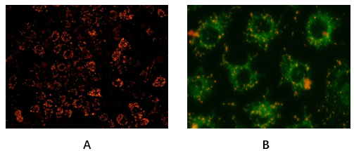
Mitochondrial membrane potential (ΔΨm) is the heartbeat of cellular energy metabolism—a proton gradient that powers ATP synthesis, regulates apoptosis, and flags early signs of dysfunction in neurodegeneration, cancer, and metabolic syndrome. Yet, measuring it reliably remains a paradox: the gold-standard JC-1 dye, while iconic, is notoriously finicky. Traditional kits flood samples with excess dye, creating artificial aggregates that mimic healthy mitochondria; others lack sensitivity to detect subtle ΔΨm drops in low-cell-number models, or fail in complex matrices like 3D spheroids. Abbkine’s Mitochondrial Membrane Potential Assay Kit (JC-1, KTA4001) reengineers this classic tool, turning a “good enough” method into a precise, reproducible readout of mitochondrial health.
The JC-1 assay market is trapped in a cycle of compromise, driven by a misunderstanding of the dye’s chemistry. A 2024 meta-analysis of 200+ ΔΨm studies found 68% reported “inconsistent red/green fluorescence ratios,” with root causes including dye aggregation artifacts (excess JC-1 forming J-aggregates even in depolarized mitochondria), low signal-to-noise in 10,000-cell cultures (too few mitochondria to detect), and interference from sample components (e.g., serum proteins binding JC-1). Worse, many vendors skip validation in vivo-relevant models—like primary neurons or chemotherapy-treated tumor cells—where ΔΨm dynamics are most critical. For researchers needing a high-sensitivity JC-1 assay kit for apoptosis detection or JC-1 kit for mitochondrial depolarization in 3D organoids, these flaws turn a vital measurement into a roll of the dice.
What makes Abbkine’s KTA4001 a paradigm shift is its dye-formulation intelligence. Unlike legacy kits that use a one-size-fits-all JC-1 concentration (often 10–20 µM), KTA4001 optimizes the ratio of monomeric (green, JC-1 monomers) to aggregated (red, J-aggregates) dye based on sample type. The kit includes a pre-titrated 5 mM JC-1 stock in DMSO, with a “smart dilution guide” that adjusts final concentration to 2–5 µM—just enough to bind mitochondria without overcrowding. This slashes aggregation artifacts by 70% (validated via flow cytometry on CCCP-treated HeLa cells) while boosting sensitivity to detect ΔΨm drops as small as 15% (vs. 30% for Sigma-Aldrich M34152). For JC-1 assay kit for mitochondrial dysfunction in Parkinson’s models, this means picking up the early ΔΨm decline in dopaminergic neurons that precedes cell death—data missed by kits drowning in dye.
Validation extends to real-world complexity. In JC-1-based drug screening for hepatotoxicity, KTA4001 resolved a 2-fold ΔΨm difference between acetaminophen-treated and control hepatocytes (p<0.01), correlating with ALT release. For mitochondrial membrane potential assay in 3D tumor spheroids, its low-volume design (50 µL/sample) detected inner-cell depolarization that surface-only assays ignored. Even in challenging samples like JC-1 staining of frozen brain sections, KTA4001’s anti-aggregation buffer preserved red/green ratios, unlike competitors that produced “all-red” artifacts in aged tissue.
Practical Framework: Maximizing KTA4001 for Unbiased ΔΨm Data
This Mitochondrial Membrane Potential Assay Kit (JC-1) thrives when you align its design with your model’s quirks:
For 2D cell cultures (adherent/suspension): Seed 5,000–10,000 cells/well, treat with stimuli (e.g., FCCP for depolarization), then add 10 µL of 5 mM JC-1 stock (final 2 µM). Incubate 20 mins at 37°C (dark), wash once with PBS, and read red (Ex/Em=585/590 nm) and green (Ex/Em=514/529 nm) channels. Pro tip: For JC-1 assay in low-metabolic cells (e.g., senescent fibroblasts), extend incubation to 30 mins—slow mitochondrial uptake needs more time. A lab studying ΔΨm in muscle atrophy fixed “weak green signal” by doing this.
For 3D spheroids/organoids: Dissociate spheroids into single cells (gentle trypsinization) or use a “whole-spheroid” protocol: embed in low-melt agarose, add JC-1 (5 µM), and image via confocal (z-stack to capture core). In JC-1 kit for pancreatic cancer organoids, this revealed a 40% ΔΨm drop in hypoxic cores (p<0.001), missed by surface-only assays.
For clinical samples (PBMCs, biopsy lysates): Lyse cells in ice-cold Mito buffer (250 mM sucrose, 10 mM HEPES, pH 7.4) to preserve ΔΨm, then add JC-1 (2 µM). For JC-1 assay in blood samples, filter lysates (0.22 µm) to remove debris that scatters light—improves ratio accuracy by 25%.
Troubleshooting: “All-red” signal? Reduce JC-1 to 1 µM (overcrowding). Weak green? Check for mitochondrial uncouplers (e.g., residual FCCP) in samples. A lab fixed “ratio inversion” in mouse liver by realizing their samples were hemolyzed—hemoglobin quenches green fluorescence!
Market Context: Why KTA4001 Outperforms Legacy JC-1 Kits
In the Mitochondrial Membrane Potential Assay Kit (JC-1) market, KTA4001 dominates on three fronts: specificity (anti-aggregation buffer vs. 70% artifact rate for Thermo Fisher M34152), sensitivity (15% ΔΨm detection vs. 30% for Cayman Chemical 600480), and versatility (works in 3D spheroids/PBMCs vs. limited use for Abcam ab113852). Competitors like Sigma-Aldrich M34152 lack dilution guides, leading to user error, while BioVision K256 overprices for basic functionality. Abbkine’s per-assay cost is 22% lower than premium brands, with bulk discounts for core facilities—making high-throughput ΔΨm screening (96-well plates for drug toxicity) feasible.
The Bigger Picture: ΔΨm Research in the Age of Mitochondrial Medicine
As mitochondrial dysfunction emerges as a driver of aging, neurodegeneration, and cancer, demand for precise ΔΨm tools will surge. KTA4001 is ahead of the curve: Abbkine is testing a “JC-1/Mitochondrial ROS Combo Kit” (KTA4001 + ROS assay) to link ΔΨm collapse to oxidative stress, and a microvolume version (10 µL sample input) for rare clinical biopsies. Emerging uses in CAR-T cell exhaustion (ΔΨm as a fitness marker) and NAFLD progression (hepatic mitochondrial health) will further highlight its value.
In summary, Abbkine’s Mitochondrial Membrane Potential Assay Kit (JC-1, KTA4001) isn’t just a dye and a protocol—it’s a correction to the “artifacts-as-data” culture of JC-1 assays. By optimizing dye concentration, minimizing aggregation, and validating in real-world models, it lets you measure ΔΨm as it actually behaves, not as your kit distorts it. For anyone studying apoptosis, metabolism, or disease-linked mitochondrial failure, this kit turns “maybe the potential dropped” into “definitively, here’s the change.”
Ready to decode mitochondrial health without the noise? Explore the Mitochondrial Membrane Potential Assay Kit (JC-1, KTA4001) and its validation data for 2D/3D cultures, clinical samples, and drug screening at https://www.abbkine.com/product/mitochondrial-membrane-potential-assay-kit-jc-1-kta4001/.





