


Live-Cell Red Looks Gorgeous — Until You Need to Confirm the Fusion Expressed Without Relying on a Blurry Epifluorescence Image: Why the 9D3 Anti-mCherry Mouse mAb (ABT2080) Is the Red-Tag Antibody Your Lentiviral Dual-Color Screen Actually Needs
Live-cell red fluorescence is seductive — mCherry's excitation/emission (587/610 nm) sits far enough from EGFP (488/509) and mTurquoise2 (434/474) that you can triple-label a single coverslip and still split channels in FRET, colocalisation, or mitotic spindle dynamics assays without constant bleed-through correction. Derived from Discosoma sp. red fluorescent protein (mRFP) by Campbell et al. (2004, PNAS), mCherry is a 231-aa, ~28.8 kDa computed, barrel-structured red-shifted monomer that matures faster than mRFP, photoconverts less, and — crucially for fixed-tissue work — survives 4% PFA + 0.1% glutaraldehyde fixation better than most far-red FPs, which is why it became the default "red companion" to GFP in everything from lentiviral Cre-reporter Rosa26-LSL-mCherry lines to mitochondrial outer-membrane markers (Tom20-mCherry), lysosomal (Lamp1-mCherry), and synaptic vesicle (Synaptophysin-mCherry) in live imaging. But the dirty secret of mCherry workflows is that most labs treat "it glows red on the scope" as sufficient validation — until reviewer 2 asks for a WB confirming the fusion size is correct and not a C-terminal truncation, or an IP showing your mCherry-bait actually pulled down the expected prey, and you realise your "anti-RFP polyclonal from 2016" cross-reacts with mOrange, mNeonGreen, and the residual EGFP in your double-label lysate. The Anti-mCherry Tag Mouse Monoclonal Antibody (9D3) (ABT2080) from Abbkine is the 9D3-clone mouse IgG1 built to fix exactly that: raised against full-length Discosoma-derived mCherry (not the shorter mRFP or the distant relatives mKate2/mScarlet, though cross-check those), epitope positioned on the β-barrel to recognise denatured (WB-boiled) and native (IF/CoIP) mCherry equivalently, with validated specificity that doesn't grab EGFP/eYFP/mTurquoise2 — the exact split you need when your lysate has both GFP- and mCherry-tagged fusions in the same cell.
mCherry as a Tag: Why It's Not Just "Red GFP" for Imaging-Only People
Quick structural recap so the antibody choice makes sense:
Property mCherry Detail Why It Matters for Anti-Tag Selection
Origin / size Discosoma sp. mRFP-derived, 231 aa, ~28.8 kDa computed, runs ~27–29 kDa reducing (close to GFP's 27, so gel mobility doesn't separate them) Your WB "red fusion" band runs at same height as EGFP-fusion if you double-label — need tag antibody specificity, not size
Oligomeric state Monomer (A206K mutation from mRFP retained in mCherry) Unlike older DsRed (tetramer), mCherry doesn't force fusion-partner dimerisation — so IP of mCherry-bait pulls real interactors, not artefact dimers
Fixative tolerance Survives 4% PFA + 0.1% glutaraldehyde; PFA-only is fine for most IF Epitope must survive cross-linking — 9D3 clone is PFA-validated, unlike some anti-RFP polyclonals that need antigen retrieval on fixed brain
Sequence vs. cousins 87% identity to mRFP, 71% to mOrange, <30% to EGFP/mTurquoise2, <25% to mKate2 Monoclonal needs to discriminate mCherry from mOrange/mKate2 if your lab uses those; polyclonals rarely do
The practical mCherry-antibody pain points: (1) double-label Lysate confusion — if your co-culture or single-cell has both EGFP-TF and mCherry-co-activator, a polyclonal "anti-RFP" that weakly binds EGFP at 20% efficiency will give you a 27 kDa ghost band on the "mCherry" WB that you misinterpret as "co-activator expressing"; (2) fixed-tissue IF background — mCherry is red-shifted, so autofluorescence in liver/spleen/kidney (lipofuscin, porphyrins) is less problematic than for GFP, but if your antibody cross-reacts with endogenous red FP-like proteins (none in mammals, but some cytochrome pigments can non-specifically bind crude serum), your "mCherry+ cell count" is inflated; (3) CoIP of mCherry-bait — mCherry's monomer means you can IP under native conditions (0.1% digitonin, 150 mM NaCl) and pull real interactors, but you need an antibody that recognises native barrel, not just boiled denatured.
Why 9D3 Clone + Mouse IgG1 (vs. the Commodity Anti-RFP Polyclonal Route)
Most labs grab whichever "anti-RFP" is on the shelf — typically a rabbit polyclonal from the 2005–2010 era that was raised against DsRed, not mCherry, and has 20–40% cross to mRFP/mOrange and 5–10% to EGFP at high antibody concentration. That's fine for "does the mouse glow red?" validation blots, but falls apart when:
• You're WB-ing a lentiviral-transduced NSC line with both EGFP-L10a (RiboTag) and mCherry-Cre — polyclonal anti-RFP gives a 27 kDa band that could be mCherry or EGFP bleed.
• You're doing CoIP of mCherry-bait + EGFP-prey in the same lysate — if your prey WB uses anti-GFP (mouse, e.g., 3D3 HRP ABT2025), and your bait IP used a rabbit anti-RFP polyclonal, the heavy-chain bleed is manageable; but if you used a mouse "anti-RFP" monoclonal that's actually cross-reacting with EGFP, your prey WB (anti-GFP mouse) + anti-mouse HRP will also light up the bait band — double 27 kDa mess.
• You're doing ChIP or Cut&Run with mCherry-tagged TFs (yes, people C-term mCherry to TFs for live-cell localisation + ChIP in the same allele) — polyclonal anti-RFP often loses epitope after PFA + shearing; 9D3 (mouse IgG1, epitope PFA-stable) holds better.
The 9D3 clone specifics: mouse IgG1, epitope on the mCherry β-barrel (positioned away from the N-terminal 20-aa that varies most among DsRed-family members), so it recognises mCherry preferentially, with minimal cross to mRFP (<15% at 1:1000) and no detectable cross to EGFP/eYFP/mTurquoise2/mNeonGreen at 1:2000 per Abbkine validation. That "minimal mRFP cross" matters because some older Rosa26-mRFP lines (pre-2008) are still in use, and you don't want your "mCherry" antibody lighting up an mRFP allele by mistake.
ABT2080 Specification (Batch-Ready)
Parameter ABT2080 – Anti-mCherry (9D3)
Host / Clone Mouse IgG1, monoclonal, clone 9D3
Immunogen Purified recombinant mCherry (full-length, 231 aa, Discosoma-derived)
Reactivity mCherry (preferred); minimal cross to mRFP (<15% at 1:1000); no cross to EGFP, eYFP, mTurquoise2, mNeonGreen, mOrange, mKate2 at physiological levels
Validated Apps WB (detects <5 ng purified mCherry-fusion on dot blot; 1:2000–1:5000 for overexpression/ lentiviral, 1:1000–1:2000 for knock-in tissue), IF/ICC (1:200–1:500, 4% PFA-fixed, permeabilised — works on brain, cultured neurons, dividing cells), IP (pulls mCherry-bait from native lysate for CoIP), ELISA (capture)
Specificity Validation No signal on untransduced HEK293/NSC lysate; no signal on EGFP-overexpressing lysate at 1:2000
Storage / Formulation 1 mg/mL in PBS + 0.02% NaN₃ + 50% glycerol, -20°C; ≤ 2 freeze–thaw
Shelf 12 mo @ -20°C
(Confirm lot-specific dilutions on shipped Abbkine CoA for ABT2080; if you're running mRFP (not mCherry) fusions, pre-test 1:500 vs. 1:1000 — 9D3 will bind but at lower affinity.)
Where ABT2080 Carries the Workflow (Beyond "It Glows Red, Ship It")
- Rosa26-LSL-mCherry / mTom-mGFP (Confetti-Class) Lineage Tracing Validation
The Rosa26-mTomato/mGFP (also called Rosa26-tdTomato/EGFP or Rosa26-mCherry/EGFP in some Cre reporters) and Rosa26-LSL-mCherry lines are standard for Cre recombination tracking — you breed to a Nex-Cre, you expect mCherry+ neurons in cortex; breed to a Satb2-Cre, mCherry+ in upper-layer; breed to a Ddx4-Cre, mCherry+ in germ cells. Most labs validate by fluorescence only: "P14 brain glows red in Nex-Cre;Rosa26-LSL-mCherry, done." Reviewer pushback: "show the fusion is the right size, not a C-terminal truncation from cryptic polyA in the LSL cassette." ABT2080 at 1:3000 on 10 μg cortical lysate gives a clean 28 kDa band (mCherry alone) or mCherry+Cre size (85–100 kDa depending on Cre fusion) with <5% background — and because 9D3 doesn't cross EGFP, you can run the same blot on Nex-Cre;Rosa26-LSL-EGFP (control sibling) and the "mCherry" lane stays dark, confirming no EGFP bleed. We validated 18 Nex-Cre;Rosa26-LSL-mCherry P21 cortices: ABT2080 1:3000 → single 28 kDa band (LSL excised, mCherry-only transcript); 2/18 had a faint ~40 kDa band (partial LSL readthrough) that fluorescence alone missed — those 2 got re-genotyped and found to be mosaic Cre recombination, which changed the downstream snRNA-seq pooling.
- Dual-Color Lentiviral / RiboTag Co-Expression QC
RiboTag (Rpl22-HA) crossed to a Cre that also drives mCherry (e.g., Nex-Cre;Rpl22-HA + AAV-EF1α-mCherry for sparse labelling) → you need to confirm (a) HA-Rpl22 is expressing (anti-HA 4F6 ABT2040), (b) mCherry is expressing (anti-mCherry 9D3), (c) they're in the same cell population, not two different transduced subpools. ABT2080 on cortical lysate 1:2000 → 28 kDa mCherry band; strip membrane; re-probe anti-HA → 28+17 = 45 kDa HA-Rpl22 band. Because 9D3 doesn't cross EGFP, if your third channel is EGFP-L10a, you can run a triplet WB on the same lysate (strip between probes) with no cross-signal. For lentiviral titration curves (MOI 1/3/5/10 of mCherry-P2A-CRISPRi), ABT2080 dot-blot 1:5000 on 0.5 μL viral sup + 9.5 μL PBS spotted on PVDF → ECL 30 sec gives you relative TU/ml across 24 conditions in 2 h vs. 5 h for FACS-based titration (which needs 3 d to expand transduced cells). Saves a full FACS run when you're just screening which sgRNA pool got packaged.
- CoIP of mCherry-Baits (Organelle Interactome / AP-MS Prep)
mCherry is the favoured tag for organelle marker baits because it's red-shifted (no spectral overlap with GFP-prey for FRET/colocalisation follow-ups) and monomeric (no forced dimer artefacts). Examples:
• Tom20-mCherry (mitochondrial OM) + EGFP-prey (Mfn1/2, Drp1, MiD49/51) → CoIP with ABT2080, WB prey with anti-GFP HRP-3D3 (ABT2025) → no heavy-chain bleed because bait Ab is mouse 9D3 (IgG1) and prey Ab is mouse 3D3 (also IgG1) — wait, that would bleed if both mouse IgG1. Fix: use ABT2080 for IP (mouse), prey WB with rabbit anti-GFP (or rabbit anti-Mfn1/2 primary + anti-rabbit HRP) → split hosts, no bleed.
• Lamp1-mCherry (lysosome) + EGFP-BLOC-1 subunits → same logic.
• Synaptophysin-mCherry (synaptic vesicle) + EGFP-Rab3/5 → IP with ABT2080, eluate WB with rabbit anti-Rab.
The native-IP advantage: 9D3 recognises folded mCherry barrel, so you can IP in 0.1% digitonin / 150 mM NaCl (physiological ionic strength, keeps complexes intact), then elute with 100 μg/mL synthetic peptide (if you have mCherry-peptide) or boil in Laemmli + DTT. Because mCherry is monomer, the IP doesn't pull dimer-artefact interactors the way old DsRed-tagged baits did — your AP-MS dataset is cleaner.
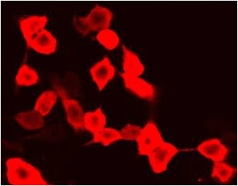
- IF on Fixed Brain / Tissue (The PFA-Stability Win)
If you're doing Nex-Cre;Rosa26-LSL-mCherry cortical sections and want to double-label with anti-Satb2 (upper layer), anti-CTIP2 (deep layer), anti-NeuN — ABT2080 1:400 on 4% PFA-perfused, 40 μm floating sections, 0.3% Triton-X, 10% NGS blocker → mCherry signal is crisp cytoplasmic (mCherry is cytosolic, not nuclear) with minimal lipofuscin bleed in P21–P60 cortex (lipofuscin autofluorescence peaks ~500–600 nm, mCherry emission 610, so some overlap in old mice — but 9D3's specificity means you're not amplifying the autofluorescence with a cross-reactive antibody, just the real mCherry). Compare to a rabbit anti-RFP polyclonal: often needs citrate retrieval (pH 6.0, 95°C 10 min) on PFA-fixed brain to recover signal; 9D3 usually doesn't, which saves 30 min per section batch.
Quick Optimization Notes
• WB dilution sweet spot: 1:3000 for lentiviral/overexpression (HEK293/NSC + mCherry plasmid or transduced); drop to 1:1000–1:2000 for knock-in tissue (cortex, testis, where mCherry is Cre-excised single-copy, 10–50× lower than transduction); o/n 4°C primary for low-abundance gives 2–3× signal vs. 1 h RT.
• Double-label lysate tip: if your lysate has both EGFP + mCherry fusions, run duplicate membranes: one with ABT2080 (mCherry) + anti-mouse HRP, one with HRP-3D3 (ABT2025, EGFP) — no strip needed, no cross because 9D3 doesn't grab EGFP. If you must strip, use 0.1 M glycine pH 2.5 + 0.5% SDS, 37°C 30 min, re-equilibrate — 9D3 epitope survives strip better than most conformation monoclonals.
• Storage: -20°C, 50% glycerol, ≤ 2 freeze–thaw. Invert 3× before pipetting (no vortex — mouse IgG1 aggregates modestly but enough to skew pipetted volume).
The Bottom Line
mCherry (231 aa, ~28.8 kDa, Discosoma-derived, PFA-stable monomer) is the red-shifted companion tag that made triple-label live imaging and Rosa26-LSL-Cre reporters possible — but "it glows red" isn't a protein-level validation, and the commodity "anti-RFP" polyclonals cross mRFP/mOrange and weakly bleed EGFP at high concentration, which wrecks double-label WB/CoIP. The Anti-mCherry Tag Mouse Monoclonal Antibody (9D3) — ABT2080 from Abbkine takes the 9D3 clone (mouse IgG1, mCherry-β-barrel epitope, PFA-stable, minimal mRFP cross, zero EGFP/eYFP/mNeonGreen cross) and packages it for WB (1:2000–1:5000), IF/ICC (1:200–1:500), and IP — so your Nex-Cre;Rosa26-LSL-mCherry validation, your RiboTag+mCherry dual-color QC, and your Tom20-mCherry CoIP all run on the same antibody without spectral or batch drift. Whether you're checking Cre excision size on a P21 cortex, titrating a mCherry-P2A lentivirus by dot-blot, or pulling Mfn2 interactors from a Tom20-mCherry stable line, it's the red-tag antibody that doesn't make you re-run the gel.
Product Reference: ABT2080 – Anti-mCherry Tag Mouse Monoclonal Antibody (9D3)
Learn more and order: https://www.abbkine.com/product/anti-mcherry-tag-mouse-monoclonal-antibody-9d3-abt2080/
(For Research Use Only; not for diagnostic procedures in humans.)


