| Product name | Anti-mCherry Tag Mouse Monoclonal Antibody (9D3) |
| Immunogen | Recombinant Protein |
| Host | Mouse |
| Reactivity | Mammals, Bacteria |
| Applications | IF, WB |
| Applications notes | Optimal working dilutions should be determined experimentally by the investigator. Suggested starting dilutions are as follows: WB (1:5000), IF (1:200). |
| Clonality | Monoclonal |
| Preparation method | The antibody was affinity-purified from mouse ascites by affinity-chromatography using specific immunogen |
| Alternative | mCherry; mCherry tag |
| Formulation | Liquid solution |
| Storage buffer | Liquid in PBS, pH 7.4, containing 0.02% Sodium Azide as preservative and 50% Glycerol. |
| Storage instructions | Stable for one year at -20°C from date of shipment. For maximum recovery of product, centrifuge the original vial after thawing and prior to removing the cap. Aliquot to avoid repeated freezing and thawing. |
| Shipping | Gel pack with blue ice. |
| Precautions | The product listed herein is for research use only and is not intended for use in human or clinical diagnosis. Suggested applications of our products are not recommendations to use our products in violation of any patent or as a license. We cannot be responsible for patent infringements or other violations that may occur with the use of this product. |
| Background | mCherry is a fluorophore (a fluorescent protein) used in biotechnology as a tracer to follow the flow of fluids, as a marker when tagged to molecules and cell components. mCherry, derived from a protein isolated from Discosoma sp., is a 28.8kD monomeric fluorescent construct with peak absorption/emission at 587 nm and 610 nm. mCherry is sometimes preferred to other fluorophores due to its colour, as well as its photostability compared to other monomeric fluorophores. |
| Alternative | mCherry; mCherry tag |
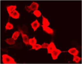
Fig.1. Immunofluorescence staining (1:200) of mCherry fusion protein in 293 cells with red.
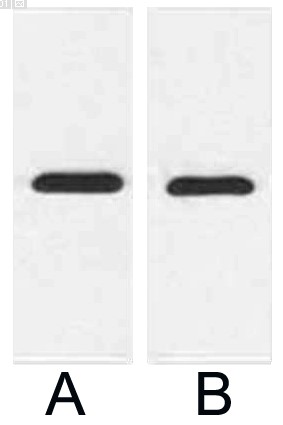
Fig.2. Western blot analysis of 1ug mCherry fusion protein with Anti-mCherry Tag Mouse Monoclonal Antibody (9D3) in 1:2000 (lane A) and 1:5000 (lane B) dilutions.
Author:Meng, Li, et al. Publication name:Nature Communications IF:15.7
Author:Zuo, Peng, et al. Publication name:Nature Communications IF:15.7
Author:Du, Linying, et al. Publication name:New Phytologist IF:8.1
Author:H Gao, Y Guo, M Ren, L Tang, W Gao, S Tian, ... Publication name:Redox biology IF:6.9
Author:R Cai, P Bai, M Quan, Y Ding, W Wei, C Liu, ... Publication name:Stem Cells IF:6.4
Author:Zhang F, Zhong R, Li S, et al Publication name:Front Aging Neurosci IF:4
Author:Lai Y, Fan L, Zhao Y, et al Publication name:Int J Oncol IF:4
Author:Oumei Cheng, Xiaoyan Tian, et al Publication name:Oncotarget IF:4
Author:Liu, L, et al Publication name:Scientific Reports 5(2015):14474 IF:4
Author:Dong, Chaoran, et al Publication name:Scientific Reports 6(2016): 36860 IF:4
Author:Zhang L, Song J, Bai T, et al Publication name:Sci Rep IF:4
Author:Xia Zhong, Qian-Qian Wang, et al Publication name:Scientific Reports IF:4
Author:Tan R J, Sun H Q, Zhang W, et al Publication name:Helicobacter IF:3
Author:Yan, Ruyu, et al Publication name:Inflammation Research 64 IF:3
Author:Chen Q, Wang K, Jiang D, et al Publication name:Neurochemical research IF:3
Author:Xu Y, Meng C, Liu G, et al Publication name:The Journal of the American Society of Anesthesiologists IF:3
Author:Zuo Y C, Xiong N X, Shen J Y, et al Publication name:Neurochemical Research IF:3
Author:Lv L, Liu H G, Dong S Y, et al Publication name:Tumor Biology IF:3
Author:Geng, Xin, et al Publication name:World Neurosurgery 100 (2017): 407-416 IF:2
Author:Fu H D, Wang B K, Wan Z Q, et al Publication name:Journal of Molecular Histology IF:2
You must be logged in to post a review.


Reviews
There are no reviews yet.