




Your “Loading Control” Isn’t Just a Housekeeping Band—It’s the Silent Judge of Every Western & IHC You Publish. Here’s Why ABP0056 Is the Rabbit Polyclonal Actin Antibody That Stops Reviewer Side-Eye
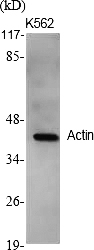
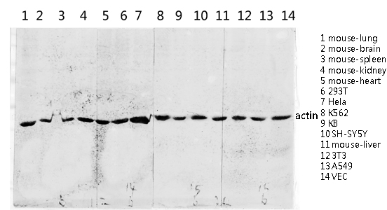
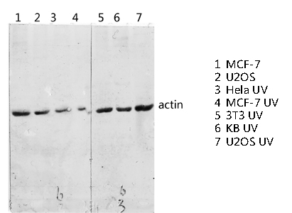
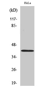
If you’ve ever had a methods-minded reviewer ask “How was the actin/β-actin antibody validated for this tissue and detection system?”—and you realized your “actin” lane is a smudge at 42 kDa flanked by weird cross-reactivity—you already know the dirty truth: the most-used antibody in biology is also the one most often chosen on autopilot. The problem isn’t that actin moves; it’s that not every anti-actin reagent is built to survive the jump from a 1:500 Western blot to paraffin IHC-P without falling apart.
Actin Is “Boring” Only on the Surface—and That’s Why Specificity Still Matters
β-Actin (ACTB) is the canonical cytoplasmic, non-muscle isoform that forms the core of the cytoskeleton and participates in cell motility, structural integrity, and even nuclear architecture. Because actin is hyper-conserved across Human–Mouse–Rat, a well-designed antibody can be genuinely cross-reactive in the right way—detecting the endogenous, ~42–43 kDa β-actin that you actually want as a normalization/architecture anchor—without dragging in a carnival of non-specific bands that make your densitometry look generous.
The mistake most labs make is treating actin like a “any-rabbit-anti-actin will do” checkbox. Once you move into formal-fixed, paraffin-embedded (FFPE) IHC-P or you push a blot to 1:2,000+ for a clean, publication-ready figure, the difference between a serum-derived polyclonal soup and an affinity-conscious, epitope-informed rabbit IgG becomes very visible, very fast.
Meet ABP0056 — Rabbit Polyclonal Actin Antibody (ABBKINE, ABP0056)
This isn’t a “generic actin” tag sourced from whoever was cheapest that quarter. ABP0056 is a rabbit polyclonal IgG raised against a synthesized peptide derived from the C-terminal region of human Actin (AA range ~300–380), and it’s formulated the way a serious core or a publication-focused lab actually needs it:
Parameter ABP0056 (Actin Polyclonal Antibody)
Brand / Cat# ABBKINE • ABP0056
Target Actin / β-Actin (ACTB) — cytoplasmic, non-muscle isoform (highly conserved)
Immunogen Synthesized peptide derived from C-terminal region of human Actin (AA 300–380)
Host / Isotype Rabbit · IgG · Polyclonal
Reactivity Human, Mouse, Rat (C-terminal epitope is extremely well conserved)
Validated apps WB, IHC-P, ELISA (others not yet tested)
Suggested starting dilutions WB 1:500–1:2,000 · IHC-P 1:100–1:300 · ELISA 1:10,000
Form / Concentration Liquid · 1 mg/mL
Storage buffer PBS, pH 7.4 containing 50% glycerol, 0.5% BSA, 0.02% Sodium Azide (NaN₃)
Storage / Stability –20°C; stable 1 year from shipment date; centrifuge original vial after thawing/before opening; aliquot to avoid repeated freeze–thaw
Shipping Gel pack with blue ice
Use statement For research use only; not for human/clinical diagnostic use
Two things jump out immediately to anyone who’s been burned before:
- It’s 1 mg/mL in a glycerol/BSA/NaN₃ PBS system, which protects the IgG during freeze–store and reduces surface adsorption losses when you work at high dilutions.
- The C-terminal synthetic-peptide immunogen is a deliberate choice: it targets a region that remains strongly conserved across species, which is exactly why the same vial can serve H/M/R Westerns and IHC-P without rewriting your entire protocol.
What Changes in Your Figures When the Loading Control Behaves
① Your Western densitometry stops needing apologies.
Push ABP0056 to 1:1,000–2,000 on a clean transfer (PVDF/nitrocellulose), and you typically get a sharp ~42–43 kDa band with the signal-to-noise that lets you normalize lanes confidently—not “close enough.” Add a positive control lane (HeLa/293T/any robustly expressing β-actin lysate) once, and you’ve got a visual anchor that reviewers like.
② Your IHC-P stops looking like “tan background with attitude.”
Used at 1:100–1:300, this class of anti-actin polyclonal can highlight cytoplasmic/cytoskeletal continuity in FFPE sections when paired with a proper antigen retrieval (citrate pH 6.0 or Tris-EDTA pH 9.0, per your lab’s epoch) and a clean HRP/DAB or fluorescent secondary chain. It’s not a replacement for a pathway-antibody—it’s the internal sanity check that your morphology and your lysis aren’t lying to you.
③ One vial, multiple roles.
Because it’s also validated conceptually for ELISA applications (1:10,000 starting dilution noted), you can transition from lysate-format screening into coating/capture-validation workflows without hunting a different “ELISA-grade” actin reagent.
Bench-Hygiene Rules That Keep ABP0056 Performing Like Day-One
Even a premium polyclonal can be ruined by small, dumb habits—so treat it like the 1 mg/mL, glycerol-dense, NaN₃-containing reagent it is:
• Centrifuge briefly before first opening/after thawing so the glycerol gradient doesn’t cheat your pipetting.
• Aliquot early (e.g., 5–10 µL tubes). The buffer tolerates –20°C well, but repeat freeze–thaw is the fast track to aggregation and higher background.
• Mind the NaN₃ in WB/IF: if you’re going HRP-secondary, give the membrane thorough TBST washes after the primary step to avoid residual azide inhibiting peroxidase.
• For WB dilution scouting, start 1:1,000 on a strong lysate; most labs squeeze very clean bands even at 1:2,000.
• For IHC-P, run your standard retrieval and a 1:100 → 1:200 → 1:300 titration on a single test slide before committing a cohort; the difference in cytoplasmic crispness is usually decisive around that range.
Where ABP0056 Earns Its Spot in Real Papers (Not Just “Loading Control” Rituals)
Context Why a solid anti-β-Actin (C-terminal, rabbit pAb) matters
Classic WB normalization (treated vs. control in cell lines / tissues) Reproducible ~42–43 kDa band at 1:1,000–2,000 reduces the “our actin looks different today” problem
FFPE IHC-P morphology checks (tissue architecture, cytoplasmic continuity, necrosis edges) Acts as a quick global protein preservation readout next to your pathway marker
CRISPR/RNAi validation & stable line panels When you’re knocking *near-ubiquitous genes, actin confirms the lysate itself isn’t systematically degrading
Multi-condition toxicology / drug-response studies A dependable actin anchor makes your % of control honest across 8–12 treatment arms
ELISA / coating-validation pipelines (where actin leakage/tracked as background) A validated polyclonal with a suggested 1:10,000 ELISA dilution range is a tidy tool to have in the same box
A Drop-In Methods-Line You Can Borrow
β-Actin was detected with a rabbit polyclonal anti-Actin antibody (ABP0056, ABBKINE; immunogen: C-terminal peptide of human Actin, AA 300–380) at 1:1,000–2,000 for Western blot (observed ~42–43 kDa) and 1:200 for IHC-P on FFPE sections per the manufacturer’s recommendations, followed by HRP-conjugated anti-rabbit IgG and ECL/DAB development.
Explore the ABBKINE Actin Polyclonal Antibody (ABP0056) product page here:
🔗 https://www.abbkine.com/product/actin-polyclonal-antibody-abp0056/
(For research use only. Not for human or clinical diagnostic use. Handle NaN₃ with standard lab precautions; aliquot to avoid freeze–thaw; centrifuge before use.)





