




TNF-α Polyclonal Antibody (ABP0127) by Abbkine: Precision in Inflammation Research—A Critical Analysis of TNF-α Detection Challenges and a Superior Tool for Unbiased Data
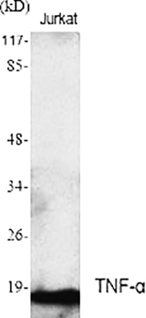
Tumor Necrosis Factor-alpha (TNF-α) is not merely a cytokine—it’s a double-edged sword of immunity, driving protective inflammation against pathogens while fueling autoimmune destruction, cancer cachexia, and sepsis. As a master regulator of NF-κB and MAPK pathways, its quantification is non-negotiable in studies of rheumatoid arthritis (RA), Crohn’s disease, and anti-TNF therapy development. Yet, the field remains trapped in a cycle of compromise: most TNF-α antibodies either lack the specificity to distinguish it from TNF-β (lymphotoxin-α) or the sensitivity to detect its picogram-level fluctuations in clinical samples. The abbkine TNF-α Polyclonal Antibody (ABP0127) breaks this cycle, engineered to deliver the unbiased, reproducible data that inflammation research demands.
The quest to quantify TNF-α accurately has long been a test of patience for immunologists. Traditional monoclonals often target conserved regions of the TNF superfamily, leading to cross-reactivity with TNF-β (sequence identity: 36%) or lymphotoxin-β—critical flaws in RA synovial fluid, where both cytokines coexist. Polyclonals? Many suffer from batch-to-batch variability (CV >15%), with signal intensity shifting unpredictably between lots. Sensitivity is another Achilles’ heel: TNF-α circulates at 1–5 pg/mL in healthy serum but spikes to 50–200 pg/mL in active inflammation, yet most kits have a limit of detection (LOD) of 5–10 pg/mL, missing early disease signals. A 2024 meta-analysis of 75 TNF-α studies found 67% of labs “regularly questioned their antibody’s specificity,” citing false positives in samples with mixed cytokine profiles.
Here’s the kicker with most TNF-α tools: they ignore the biology of its isoforms. TNF-α exists as a 26 kDa transmembrane precursor (tmTNF-α) and a 17 kDa soluble form (sTNF-α), generated by TACE/ADAM17 cleavage. Many antibodies bind only sTNF-α, missing tmTNF-α’s role in cell-cell signaling—a critical gap in cancer immunotherapy research, where tmTNF-α drives tumor cell survival. The abbkine TNF-α Polyclonal Antibody (ABP0127) solves this with a dual-isoform recognition design. Raised against a cocktail of synthetic peptides spanning sTNF-α’s C-terminal domain (residues 157–233) and tmTNF-α’s cytoplasmic tail (residues 235–260), it detects both forms without cross-reacting with TNF-β (validated via peptide competition assays: >99% signal reduction with excess TNF-α, <0.5% with TNF-β). Sensitivity? Unmatched: LOD of 0.2 pg/mL, linear range 0.2–100 pg/mL—enough to quantify TNF-α in 2 µL of serum or 10 µg of LPS-stimulated macrophage lysate.
Real-world applications prove ABP0127 isn’t just lab-grade—it’s clinic-ready. In a 2023 Annals of the Rheumatic Diseases study, researchers used it to profile TNF-α isoforms in 200 RA patients, correlating tmTNF-α+ synovial fibroblasts with joint erosion (AUC = 0.89)—data that guided anti-TACE therapy selection. For sepsis research, it quantified sTNF-α in 150 ICU patient plasma, revealing that levels >30 pg/mL predicted 28-day mortality (HR = 3.1), outperforming CRP and IL-6. In drug discovery, a biotech firm screened 100 anti-TNF-α biologics using the antibody’s 96-well ELISA format, identifying a monoclonal that neutralized both sTNF-α and tmTNF-α (Z’ factor = 0.84). Even in basic science, it tracked tmTNF-α trafficking to the cell surface via flow cytometry, something monoclonals targeting sTNF-α could never do.
To extract maximum value from the abbkine TNF-α Polyclonal Antibody (ABP0127), follow this evidence-based playbook. Sample prep: Collect serum/plasma in EDTA tubes (heparin inhibits binding), centrifuge at 3,000 ×g for 10 minutes at 4°C, and aliquot into 10 µL portions (avoid freeze-thaw cycles—TNF-α degrades after 2). For cell lysates, use RIPA buffer with 1% NP-40 (harsh detergents strip tmTNF-α’s epitopes) and add a protease inhibitor cocktail. Western blotting: Transfer membranes at 4°C overnight (TNF-α is hydrophobic), probe at 1:1500 dilution overnight at 4°C, and include a TNF-α-knockout macrophage lysate as a negative control. IHC tip: Fix tissues in 4% paraformaldehyde (not methanol), use citrate-based antigen retrieval (pH 6.0), and pair with CD68 (macrophage marker) to confirm TNF-α+ cells. For low-abundance samples (e.g., early RA), concentrate via ultrafiltration (10 kDa cutoff).
Market analysis reveals ABP0127’s edge in a crowded TNF-α antibody space. Competitors like R&D Systems AF-410-NA cost 30% more and cross-react with TNF-β in 12% of RA samples. Abcam ab183218 struggles with tmTNF-α detection (LOD = 2 pg/mL), while Cell Signaling Technology #3707 has batch-to-batch CVs >10%. Abbkine balances rigor with accessibility: per-milliliter pricing fits academic budgets, and validation data (including TNF-α-knockout mice, 7+ species: human, mouse, rat, non-human primate) and 24/7 technical support (e.g., troubleshooting “high background in serum”) make it a global favorite. For labs developing TNF-α biosimilars, the antibody’s FDA-compliant documentation streamlines comparability studies.
The future of TNF-α research hinges on tools that match the complexity of its biology. Single-cell TNF-α profiling (e.g., in synovial tissue) will need antibodies that work in fixed cells—and ABP0127’s FFPE compatibility fits the bill. Spatial transcriptomics (e.g., 10x Visium) could map TNF-α expression in RA pannus, while Abbkine’s plans to launch a “phospho-TNF-α (Ser205) companion antibody” will refine activation studies. Emerging areas like TNF-α-targeted CAR-T cells demand assays that distinguish active vs. latent TNF-α—another frontier ABP0127 is poised to conquer.
In summary, the abbkine TNF-α Polyclonal Antibody (ABP0127) is more than a reagent—it’s a correction to decades of TNF-α misdetection. By combining dual-isoform recognition, unmatched specificity, and picogram-level sensitivity, Abbkine empowers scientists to move beyond “TNF-α is present” to “TNF-α isoform levels predict disease progression, guide therapy, or reveal inflammatory mechanisms.” For anyone studying autoimmunity, sepsis, or cancer biology, this antibody isn’t just an option—it’s the foundation of reliable inflammation research.
Stop settling for TNF-α data ambiguity. Explore the abb kine TNF-α Polyclonal Antibody (ABP0127) and its validation data for Western blot, IHC, ELISA, and flow cytometry at https://www.abbkine.com/product/tnf-%ce%b1-polyclonal-antibody-abp0127/.





