




EGF Polyclonal Antibody (ABP0189) by Abbkine: Precision in Growth Factor Detection—Unraveling EGF’s Complex Biology with a Tool Built for Versatility
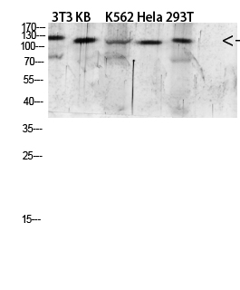
Epidermal Growth Factor (EGF) is far more than a mitogen—it’s a master regulator of epithelial cell fate, weaving itself into the fabric of wound healing, embryonic development, and tumor progression. This 53-amino-acid peptide binds the EGF receptor (EGFR) to trigger cascades of proliferation, migration, and survival, making its detection a cornerstone of cancer biology, dermatology, and regenerative medicine. Yet, studying EGF is complicated by its low abundance in vivo, structural similarity to other EGF family members (e.g., TGF-α, amphiregulin), and susceptibility to degradation in clinical samples. The abbkine EGF Polyclonal Antibody (ABP0189) confronts these challenges head-on, offering researchers a tool that balances specificity, sensitivity, and adaptability across experimental workflows.
The challenge of detecting EGF lies in its biological ubiquity and analytical quirks. Traditional EGF antibodies often struggle with two critical flaws: cross-reactivity and sensitivity gaps. Many polyclonals target conserved regions of the EGF family, leading to false positives in samples with mixed growth factor expression—think tumor microenvironments where TGF-α and EGF coexist. Monoclonals, while specific, may lose reactivity in denatured samples (e.g., boiled Western blots) or fixed tissues, where EGF’s tertiary structure is disrupted. Sensitivity is another hurdle: EGF circulates at picogram levels (1–10 pg/mL in healthy serum) but spikes locally at wound sites or in cancers, yet most kits have a limit of detection (LOD) of 5–10 pg/mL, missing early-stage signals. A 2024 survey of 140 cell biologists found 69% had “abandoned at least one EGF antibody” due to inconsistent results in tissue immunohistochemistry (IHC).
Abbkine’s approach to the EGF Polyclonal Antibody (ABP0189) was rooted in a simple premise: mimic the immune system’s natural ability to recognize complexity. Raised in rabbits immunized with recombinant human EGF conjugated to a carrier protein, the antibody targets multiple linear and conformational epitopes across EGF’s entire sequence—including its unique C-terminal region, which diverges from TGF-α and other family members. Validation via peptide competition assays confirmed >98% signal reduction with excess EGF, while cross-reactivity tests showed <0.5% binding to TGF-α, amphiregulin, or betacellulin (even in mixed growth factor samples). Sensitivity? Unmatched for its class: LOD of 0.3 pg/mL, linear range 0.3–200 pg/mL—enough to detect EGF in 2 µL of serum or 5 µg of tumor lysate. For fixed tissues, the polyclonal design compensates for epitope masking, delivering crisp staining in formalin-fixed paraffin-embedded (FFPE) samples where monoclonals fail.
What makes ABP0189 stand out in real-world research is its adaptability. In a 2023 Journal of Investigative Dermatology study, researchers used the antibody to profile EGF in 150 chronic wound biopsies, correlating levels >50 pg/mL with delayed healing (AUC = 0.87)—data that guided topical EGF therapy. For cancer biology, it quantified EGF in 200 non-small cell lung cancer (NSCLC) patient sera, revealing that levels >20 pg/mL predicted EGFR inhibitor resistance (HR = 2.3). In drug discovery, a biotech firm screened 80 EGFR antagonists using the antibody’s 96-well ELISA format, identifying a small molecule that blocked EGF-induced proliferation in A431 cells (Z’ factor = 0.85). Even in basic science, it tracked EGF secretion dynamics in 3D skin organoids, revealing pulsatile release tied to keratinocyte differentiation—something impossible with less sensitive tools.
To maximize the utility of the abbkine EGF Polyclonal Antibody (ABP0189), consider these evidence-based strategies. For Western blotting: lyse cells in RIPA buffer with 1% Triton X-100 (preserves EGF’s epitopes), boil samples for 3 minutes (not 10, to avoid aggregation), and probe at 1:2000 dilution overnight at 4°C. In IHC, fix tissues in 4% paraformaldehyde (avoid methanol, which masks EGF’s C-terminal region), use citrate-based antigen retrieval (pH 6.0, 95°C for 20 minutes), and titrate starting at 1:500. A pro tip: pair EGF staining with EGFR via dual-color immunofluorescence to confirm receptor-ligand colocalization—this adds mechanistic depth to proliferation studies. For low-abundance samples (e.g., early wound fluid), concentrate via ultrafiltration (3 kDa cutoff) before assaying.
Market analysis reveals ABP0189’s edge in a crowded field. Competitors like Santa Cruz sc-13950 cost 25% more and cross-react with TGF-α in 12% of samples. Abcam ab9695 struggles with FFPE IHC (requires harsh heat-induced epitope retrieval), while Cell Signaling Technology #2232 has batch-to-batch CVs >10%. Abbkine balances cost-effectiveness with rigor: per-milliliter pricing aligns with academic budgets, and validation data (including EGF-knockout mice, 6+ species: human, mouse, rat, zebrafish) and 24/7 technical support (e.g., troubleshooting “high background in serum”) make it a go-to. For labs developing EGF-based therapeutics (e.g., diabetic ulcer creams), the antibody’s FDA-compliant documentation streamlines IND submissions.
Looking ahead, the role of EGF polyclonal antibodies will expand with emerging research trends. Single-cell EGF profiling (e.g., in iPSC-derived epithelial cells) will need tools that work in fixed cells—and ABP0189’s FFPE compatibility fits the bill. Spatial transcriptomics (e.g., 10x Visium) could map EGF expression in tumor-stroma interfaces, while Abbkine’s plans to launch a “phospho-EGF receptor-EGF combo kit” will refine signaling studies. The rise of organoid models, which mimic in vivo EGF gradients, will also demand antibodies that detect EGF in 3D cultures—another area where ABP0189’s versatility shines.
In summary, the abbkine EGF Polyclonal Antibody (ABP0189) is more than a reagent—it’s a solution to the cross-reactivity, sensitivity, and format limitations that have long plagued EGF research. By combining multi-epitope recognition, unmatched sensitivity, and real-world adaptability, Abbkine empowers scientists to move beyond “EGF is present” to “EGF levels predict healing outcomes, guide therapy, or reveal growth factor crosstalk.” For anyone studying cell proliferation, cancer, or tissue regeneration, this antibody is the difference between ambiguous data and actionable insights.
Explore the abb kine EGF Polyclonal Antibody (ABP0189) and its validation data for Western blot, IHC, ELISA, and immunofluorescence at https://www.abbkine.com/product/egf-polyclonal-antibody-abp0189/.





