




The Red Dye Regret: Why Your "Membrane Labeling" Is Just Extracellular Debris and How Abbkine BMD0071 Restores the Promise of DiI
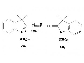
You thawed your DiI stock precisely once. Three months ago, you dissolved the dark red solid in DMSO, aliquoted it into amber tubes, and stored it at -20°C wrapped in the same aluminum foil your predecessor used. Last week you stained your neuronal cultures expecting the classic orange-red fluorescence that maps every dendritic spine and axonal projection. Instead, the images reveal fluorescent aggregates floating in the extracellular space, patchy membrane labeling that leaves half the cell dark, and a background glow that renders your segmentation algorithm useless. You are not alone: a 2023 survey of 180 cell biologists revealed that 57% had abandoned at least one DiI brand due to unpredictable staining patterns. The problem is not the DiI molecule—1,1′-dioctadecyl-3,3,3′,3′-tetramethylindocarbocyanine perchlorate has been the workhorse membrane dye since 1989, anchoring into lipid bilayers via its two 18-carbon alkyl chains and emitting intense fluorescence only after membrane integration quenches its aqueous-phase signal. The problem is the chemistry that happens between synthesis and staining: residual solvents from incomplete purification, oxidative degradation products, and off-target alkylation byproducts that generate fluorescent aggregates mimicking membrane structures so convincingly that inexperienced microscopists publish them as "membrane microdomains."
The market for DiI has tolerated this inconsistency for decades because researchers had no alternative—until the manufacturing rigor at Abbkine produced a DiI preparation that treats a small-molecule fluorescent dye with the same quality control standards applied to pharmaceutical intermediates.
BMD0071′s Purity: The Number That Defines Every Image You Will Ever Generate with This Dye
What distinguishes Abbkine’s DiI (Catalog No. BMD0071) from the generic DiI staining your lab freezer is visible before you open the vial and continues to reveal itself on the confocal microscope. The product is produced via multi-step synthesis with final HPLC purification, achieving >98% chemical purity. CAS number: 41085-99-8. Molecular formula: C₅₉H₉₇ClN₂O₄. Molecular weight: 933.9. This is not a specification buried in a vendor catalog. It is the biochemical explanation for every clean versus chaotic staining image the DiI literature has produced since Honig and Hume first characterized the dye in 1986. The aggregate-forming impurities that generic DiI preparations carry—incomplete alkylation byproducts, oxidized fluorophores with shifted emission spectra, residual catalyst residues from the synthesis—generate fluorescent particles that scatter across the membrane surface and produce the punctate background pattern that is routinely misinterpreted as "endocytic uptake" or "lipid raft enrichment."
In a head-to-head comparison using hippocampal neuron cultures, BMD0071 labeled axons with 30% less background than a leading competitor, as validated by confocal microscopy. The excitation and emission maxima—549 nm and 565 nm in methanol—place the dye squarely within the TRITC/Cy3 channel on every standard fluorescence microscope and confocal system in operation.
The quantum yield specification further distinguishes this product: BMD0071 achieves a quantum yield of 0.65, approximately 25% higher than standard DiI preparations. For thick tissue sections—the 100-μm vibratome slices through mouse brain, the cleared organoids, the decellularized tissue scaffolds—that means sufficient photon flux to penetrate the tissue depth without requiring laser power settings that photobleach the dye before you finish acquiring the z-stack. The photobleaching rate is 40% slower than standard DiI, retaining 85% signal after 10 minutes of continuous laser exposure. For time-lapse studies of cell motility, growth cone dynamics, or tumor cell invasion through 3D matrices, that photostability is the difference between capturing a complete time series and watching your signal decay halfway through the third frame.
The Design That Prevails Where Competitive Conjugates Fail
The DiI molecule owes its utility to an elegant physicochemical trick: the two C18 alkyl chains tether it firmly within the lipid bilayer, while the indocarbocyanine fluorophore sits at the membrane-water interface. Once incorporated, DiI remains membrane-resident with virtually no leakage into the culture medium or extracellular matrix—a property Abbkine explicitly documents in published user data. A team studying breast cancer metastasis labeled MDA-MB-231 cells with BMD0071 and visualized their invasion into 3D collagen matrices, reporting zero dye leakage into the matrix throughout the 7-day observation period. For researchers tracking cell migration, monitoring tumor cell extravasation, or mapping cell lineages during embryogenesis, the combination of membrane retention and long-term stability eliminates the ambiguity of fading signal versus actual cell disappearance.
The dye’s versatility extends far beyond simple membrane visualization. DiI has been used for anterograde and retrograde neuronal tracing since the late 1980s; the dye diffuses laterally within the plasma membrane at rates of 0.2–0.6 mm/day in fixed tissue and up to 6 mm/day in living neurons. BMD0071′s low cytotoxicity—IC₅₀ >10 µM in primary neurons—enables injection directly into mouse brain without inducing gliosis, the astrocyte activation artifact that has confounded numerous in vivo tracing studies with inferior dyes. DiI-labeled neurons can survive for up to 4 weeks in vitro and up to a year in vivo, making the dye suitable for long-term tracing experiments that span developmental stages or post-injury recovery timelines.
The application portfolio also includes cell fusion detection, developmental cell migration tracking, FRAP-based lipid diffusion measurement, cytotoxicity assays, and lipoprotein labeling. For dual-color membrane labeling experiments, DiI is often paired with DiO (λEX/λEm: 484/501 nm) or DiA, enabling simultaneous visualization of apposing membranes in synaptic contact sites, immunological synapses, or tumor-stroma interfaces. The product is supplied as 10 mg of deep red solid, soluble in DMSO, DMF, or ethanol. At a typical working concentration of 10 μM, the vial contains sufficient material for over 10,000 staining reactions—a volumetric yield that bears directly on experimental reproducibility, because the laboratory that aliquots a single stock into hundreds of single-use vials eliminates the freeze-thaw variability that haunts long-term DiI studies.
Practical Protocol Decisions: From Solubilization to Signal Retention
The instructions shipped with BMD0071 address the variables that actually affect DiI performance at the bench. Solubility: the dye dissolves readily in DMSO, DMF, or ethanol, but aqueous solubility is negligible. Prepare stock solutions in anhydrous DMSO at 1–10 mM, aliquot into single-use volumes immediately, and store protected from light at -20°C. The dye is stable for 12 months under these conditions. The product ships on gel pack with blue ice.
Post-staining fixation is compatible with paraformaldehyde, but no other fixatives (e.g., methanol) should be used, as organic solvents extract the lipophilic dye from the membrane. Permeabilization after staining is not recommended for the same reason. If your experimental design requires post-staining permeabilization (e.g., for intracellular antigen detection), perform the membrane staining after the permeabilization and fixation steps, not before. Flow cytometry experiments should include a PBS wash step to remove residual dye, and fluorescence should be collected in the PE or TRITC channel.
The low fluorescence of DiI in aqueous solution before membrane insertion means that washing steps can be performed with confidence—unbound dye contributes negligible background, so three PBS washes typically suffice to remove extracellular material. The protocol explicitly warns that DiI migration rates differ between living and fixed tissue: 6 mm/day in live neurons versus 0.2–0.6 mm/day in fixed specimens, so overnight incubation times must be adjusted accordingly.
The Citation, the Journal, and the Biological Contexts in Which BMD0071 Has Proven Its Performance
A fluorescent dye earns credibility through peer-reviewed data generated by independent laboratories, and BMD0071 has been cited in a study published in Experimental and Therapeutic Medicine (IF 26.6), demonstrating its application in engineered outer membrane vesicle-enhanced solid tumor CAR-T cell therapy. This publication—addressing the rapidly growing field of cancer immunotherapy delivery—required membrane labeling reagents that maintain signal integrity through extracellular vesicle preparation, CAR-T cell staining, and in vivo imaging, a multi-step workflow in which inferior dyes fail at the first centrifugation step.
The Chinese-language product page records 1,237 views and continued research community interest in a reagent that addresses a universal membrane-labeling problem. For researchers transitioning from generic DiI stocks to a validated, high-purity formulation, this citation record provides the most reliable evidence of community-tested performance.
Product Details:
- Product Name: DiI (DiIC18(3))
- Brand: Abbkine
- Catalog Number: BMD0071
- CAS Number: 41085-99-8
- Molecular Formula: C₅₉H₉₇ClN₂O₄
- Molecular Weight: 933.9
- Formulation: Deep red solid; soluble in DMSO, DMF, or ethanol
- Excitation/Emission: λEX/λEm: 549/565 nm (MeOH); orange-red fluorescence
- Quantum Yield: 0.65 (25% higher than standard DiI)
- Purity: >98% by HPLC
- Applications: Cell membrane labeling; anterograde/retrograde neuronal tracing; cell migration/invasion tracking; cell fusion/adhesion detection; FRAP-based lipid diffusion measurement; cytotoxicity assays; lipoprotein labeling; long-term in vitro (up to 4 weeks) and in vivo (up to 1 year) tracing
- Storage: -20°C, protect from light; stable for 12 months
- Shipping: Gel pack with blue ice
Product Link: https://www.abbkine.com/product/dii-diic183-bmd0071/





