


The Mitochondrial Phosphatase That Decides Between Mitophagy and Survival: Why Quantifying PGAM5 (PPM1K Family) Actually Matters for Parkin Pathways, Ischemic Injury, and Cancer Metabolism
If you've been following the mitophagy field lately, you already know the headline: PINK1 gets stranded on healthy mitochondria and vanishes, depolarized mitochondria lose ΔΨm and let PINK1 accumulate, PINK1 phosphorylates ubiquitin and Parkin, and the OMM gets tagged for autophagic clearance. But the signal that runs parallel to Parkin — and occasionally overrides it — is a Zn²⁺-dependent mitochondrial phosphatase whose name sounds like a glycolytic enzyme but whose job is 100% about life-or-death decisions at the organelle surface: PGAM5 (Serine/threonine-protein phosphatase PGAM5, mitochondrial; UniProt: Q9H0W6, Gene ID: 55276). Despite being discovered as part of the phosphoglycerate mutase family 5 fold, PGAM5 is not a glycolytic enzyme at all — it's the mitochondrial outer membrane / intermembrane space phosphatase that dephosphorylates the two most consequential mitophagy adapters (FUNDC1 and BNIP3L/NIX) and, via KEAP1, flips the NRF2 antioxidant response when mitochondria start dumping ROS. The Human PGAM5 ELISA Kit (KTE61237) from Abbkine is built to give you a quantitative, two-site sandwich ELISA readout of this pivotal phosphatase — so your "mitophagy engaged / NRF2 flipped" story rests on a calibrated ng/mL, not a ~27 kDa band you hope you normalized correctly.
PGAM5 in One Clean Pass: A Mitochondrial Phosphatase That Connects ROS → NRF2 and ΔΨm ↓ → Mitophagy
Human PGAM5 is synthesized with an N-terminal mitochondrial targeting sequence that gets cleaved after import, leaving a mature ~24–27 kDa transmembrane/anchored protein with a C-terminal PPM-type phosphatase domain that coordinates a Zn²⁺ ion instead of the classic Mg²⁺/Mn²⁺ preference. It exists in two isoform flavors from alternative translation starts:
• PGAM5L ("Long", ~287 aa) — retains a transmembrane anchor that seats it at the OMM/IMS interface, positioning the catalytic domain where it can access substrates like FUNDC1 (Ser-13/17/30), BNIP3L/NIX, and Mfn2.
• PGAM5S ("Short", ~246 aa) — truncated but still enzymatically active in many assays.
The functional circuit that made PGAM5 famous has two limbs:
Limb 1 — Mitophagy (OMM dephosphorylation)
Mitochondrial depolarization (FCCP/oligomycin+antimycin, PINK1-PRKN activation, hypoxia, mito-ROS) → calcineurin dephosphorylates PGAM5 itself → PGAM5 dephosphorylates FUNDC1 (at Ser-17 etc.) → FUNDC1's LC3-interacting region (LIR) engages → selective mitophagy. Parallel path: PGAM5 dephosphorylates BNIP3L/NIX → same net effect: autophagic clearance of the damaged unit.
Limb 2 — KEAP1 Dephosphorylation → NRF2 Activation
PGAM5 also dephosphorylates KEAP1 at Ser-104 (and possibly other sites in the BTB domain region), weakening KEAP1–CUL3 association and promoting NRF2 nuclear translocation — turning mitochondrial stress into a systemic antioxidant / detox transcriptional program.
The paradox that keeps PGAM5 interesting is: it's simultaneously pro-clearance (mitophagy) and pro-survival (NRF2). Knock it out → damaged mitochondria accumulate AND antioxidant defenses drop → bigger cell death under persistent stress. Over-activate it → excessive mitophagy → metabolic crisis in post-mitotic cells (cardiomyocytes, neurons).
Why a Sandwich ELISA for PGAM5 — And Why a Western Alone Leaves Ambiguity
PGAM5 is membrane-associated (OMM/IMS), moderately abundant only in mitochondria-rich cells, and runs at that annoying ~25–30 kDa / runs ~27 kDa region where cytosolic overflow bands and cross-reactive PGM-family proteins can haunt a blot. A two-site sandwich ELISA fixes the main reproducibility problems:
- Two independent anti-PGAM5 epitopes (pre-coated capture + biotinylated detection) → far higher specificity for the genuine phosphatase, not just "something at 27 kDa that blot-transferred."
- On-plate recombinant standard curve → OD₄₅₀ → interpolated ng/mL, so your "FCCP time course" or "PARK2 rescue" is a dataset with CVs, not a densitometry argument.
- Throughput: 96-well format means you can run multiple fractions (cytosol vs. crude mito pellet lysate), dose-responses, and genotype panels without chaining yourself to a transfer stack.
Assay Principle: KTE61237 — Sandwich ELISA
The kit follows the field-standard architecture:
- A microplate is pre-coated with a capture antibody specific for human PGAM5.
- Standards (recombinant human PGAM5) and samples (serum, plasma, tissue homogenates, cell lysates, cell culture supernatants/lysates, other biological fluids) are added; PGAM5 present binds.
- Wash → biotinylated anti-PGAM5 detection antibody (different epitope) forms sandwich.
- Streptavidin–HRP → TMB → color ∝ bound PGAM5.
- Stop → read 450 nm → interpolate unknowns from the PGAM5 standard curve.
Typical performance envelope you'll cite for this kit family:
Parameter Typical KTE61237-class specification
Target Human PGAM5 / mitochondrial Ser/Thr phosphatase (UniProt Q9H0W6, Gene ID 55276)
Format 96-well sandwich ELISA, pre-coated capture
Detection Biotin-Ab → SA-HRP → TMB, 450 nm
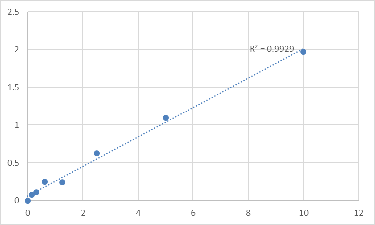
Dynamic Range 0.156 – 10 ng/mL (7-point standard)
Sensitivity / LOD ~0.05–0.10 ng/mL
Intra-Assay CV < 7–8%
Inter-Assay CV < 10–12%
Samples Tissue homogenates, cell lysates, membrane/mitochondrial fractions, other biological fluids
Assay time ~3–5 hours
(Confirm exact range, dilution scheme, and lot-specific recovery on the shipped datasheet/CoA.)
Where Quantifying PGAM5 Protein Actually Moves the Story
- Parkinsonian & PINK1–PRKN Pathway Models
PGAM5 sits just downstream of the PINK1–Parkin axis (and can also run parallel via calcineurin/FUNDC1). In MPTP/MPP⁺, rotenone, or PINK1-KO iPSC-neuron/DA-neuron models, quantifying PGAM5 protein in crude mitochondrial pellets (or OMM-enriched fractions) gives you the phosphatase pool that decides whether FUNDC1 BNIP3L are dephosphorylated — and whether the cell commits to mitophagy or inflames.
- Ischemia–Reperfusion (Heart, Kidney, Brain)
The canonical trigger: ischemic depolarization → ΔΨm ↓ → PGAM5 (calcineurin-primed) dephosphorylates FUNDC1 → mitophagy rescues some cells, but over-clearance or failed reperfusion worsens injury. Cardiomyocyte and renal proximal tubule models benefit enormously from PGAM5 quantification in fractionated lysates (expressed as ng PGAM5 / mg total protein or / mg TOM20/VDAC) so the mitophagy decision is an actual number.
- Cancer Metabolism & Chemotherapy Resistance
Damaged mitochondria in tumor cells must be managed — not eliminated too aggressively (they need energy), not ignored (ROS kills). PGAM5 levels (and the PGAM5–FUNDC1–NIX arm) modulate how tumor cells handle:
• Mitochondrial poisons (BH3 mimetics, BETi, TKIs)
• Hypoxia–reoxygenation
• Metabolic stress
Measured properly, PGAM5 becomes a therapeutic stratification marker: high PGAM5 → intact mitophagy → possibly more resistant to mito-toxic drugs; low PGAM5 → NRF2 may still save it, but mito-damage accumulates.
- PARL–PGAM5–NRF2 Crosstalk
PARL (presenilin-associated rhomboid-like protease) cleaves PGAM5's TM segment at the IMS face to generate a truncated soluble piece — the balance between intact OMM-anchored PGAM5 (mitophagy-competent) and cleaved, possibly signaling fragments is a quality-control readout in its own right. ELISA-quantified total PGAM5 in mitochondrial vs. post-mito supernatant lets you map that partitioning.
- CRISPR/AAV Validation
Editing PGAM5 (or FBXL4-related mitochondrial quality-control genes)? Don't just show "band gone." Report % PGAM5 protein remaining ± SEM from a calibrated curve, normalized to mg total protein (BCA) and an OMM marker (TOM20/VDAC), so reviewers see stoichiometry, not a gel guess.
A Minimal Prep Blueprint (Because PGAM5 Is Mitochondrial & Zn²⁺-Metal Dependent)
• Homogenize in cold sucrose/Tris buffer (e.g., 250 mM sucrose, 10 mM Tris pH 7.4, 1 mM EDTA, protease inhibitors) → differential centrifugation:
• 600–800 ×g × 10 min → discard pellet (nuclei/debris)
• 10,000–12,000 ×g × 15 min → crude mito-enriched pellet (P10) → gently lyse in 0.5–1% Triton X-100 / 150 mM NaCl + 1–5 mM DTT? (careful: DTT reduces Zn²⁺ coordination — if your assay uses Zn²⁺-dependent epitopes, keep reducing agents modest or omit pending kit guidance) + protease inhibitors
• Clarify lysed pellet again → this is your PGAM5-enriched readout
• BCA the same final lysate → express as ng PGAM5 / mg total protein or / mg TOM20.
• Warm kit reagents to RT ≥ 30 min before opening; protect TMP–TMB from light; read 450 nm promptly; run the full standard curve on every plate.
The Bottom Line
PGAM5 is the mitochondrial phosphatase that decides whether a damaged organelle gets eaten (FUNDC1/BNIP3L-driven mitophagy) or the cell calls in detox reinforcements (KEAP1/NRF2) — and when that decision flips wrong, neurons die, hearts fail, and tumors outlive their mitochondria. Measuring it as a calibrated variable instead of a "27 kDa band" changes the resolution of your entire mitophagy story. The Human Serine/threonine-protein phosphatase PGAM5, mitochondrial (PGAM5) ELISA Kit — KTE61237 from Abbkine gives you the assay architecture to do that: pre-coated capture → biotin detection → HRP–TMB → 450 nm → ng/mL, in a ~3–5 hour workflow that scales across fractions, conditions, and cohorts without a gel-stack detour.
Product Reference: KTE61237 – Human Serine/threonine-protein phosphatase PGAM5, mitochondrial (PGAM5) ELISA Kit
Learn more and order: https://www.abbkine.com/product/human-serine-threonine-protein-phosphatase-pgam5-mitochondrial-pgam5-elisa-kit-kte61237/
(For Research Use Only; not for diagnostic procedures in humans.)


