


The Cytokine That Plays Both Savior and Villain: Why TGF‑β1 Detection Demands a High‑Fidelity Polyclonal Antibody — And How Abbkine ABP52598 Delivers
If there is one pleiotropic cytokine that can make or break your experiment—and your therapeutic hypothesis—it's Transforming Growth Factor‑beta 1 (TGF‑β1). It is the archetype of context‑dependent biology: in early injury, it shuts down inflammation, drives tissue repair, and keeps epithelial barriers intact; left unopposed or reactivated chronically, it becomes the master puppeteer of fibrosis, immunosuppression, and the tumor microenvironment's "pro‑cancer" armor. Because so much of its biology hinges not just on how much TGF‑β1 is made, but on whether it is latent vs. active, and where it sits (matrix‑bound, cell‑associated, or soluble), your antibody choice is never "just a reagent"—it is the lens that decides whether you see biology or an artifact.
TGF‑β1 101: A Master Switch Disguised as a Simple Dimer
TGF‑β1 is produced as a ~390 aa precursor (pre‑pro‑TGF‑β1) that gets cleaved by furin‑like convertases to release the mature ~12.5 kDa homodimer (disulfide‑linked) and the LAP (Latency‑Associated Peptide, ~75 kDa pro‑region fragment). What leaves the cell is overwhelmingly in the small latent complex (SLC = LAP + mature TGF‑β1, non‑covalent), often covalently tethered via LAP to LTBP (Latent TGF‑β Binding Protein) in the ECM. Activation—i.e., liberating the bioactive dimer—requires mechanical force, integrin αvβ6/αvβ8, thrombospondin‑1, plasmin, acidic microenvironments, or even ionizing radiation to break LAP's grip. This elegant latency explains why total‑TGF‑β1 staining/quantification and bioactivity assays (e.g., mink lung luciferase/ALK5‑reporter) often tell different stories—and why antibody specificity, epitope accessibility, and knowledge of your sample prep are non‑negotiable.
Why a "Polyclonal" Against TGF‑β1 Is Still the Smart Default for Many Labs
Monoclonals are elegant, but a well‑made polyclonal against a carefully chosen immunogen (usually the mature dimer region or a conserved C‑terminal epitope rather than LAP's most conformation‑sensitive loops) brings practical advantages:
• Multi‑epitope recognition → better sensitivity for both free mature TGF‑β1 and LAP‑embedded forms (provided your denaturation/antigen retrieval exposes them).
• Signal robustness → often shines in IHC/ICC on FFPE, where epitope masking is the real enemy, not just affinity.
• Flexibility → one serum/affinity‑purified reagent that can span Western blot, IHC‑P, ICC/IF, and even ELISA coating/capture saves optimization time when you're pivoting between platforms.
The catch? Polyclonal quality lives or dies by specificity control: you need a supplier who validates no cross‑reactivity to TGF‑β2/TGF‑β3 at physiologically relevant ranges, and who documents species reactivity (human, mouse, rat…) and application‑level QC blots so your Figures survive peer review.
Meet ABP52598: Abbkine TGF‑β1 Polyclonal Antibody (Rabbit, Affinity‑Purified)
The TGF‑β1 Polyclonal Antibody (ABP52598) from Abbkine is positioned for researchers who want a single, affinity‑purified rabbit IgG that can bridge detect and localize TGF‑β1 across multiple contexts without forcing you into a brand‑locked detection ecosystem.
What the datasheet typically emphasizes (and why it matters to you):
• Host / Format: Rabbit polyclonal, affinity‑purified (so you're not dragging along unrelated Ig that spikes background in IHC/IF).
• Immunogen logic: Usually derived from the mature human TGF‑β1 sequence region—meaning the antibody is biased toward the biologically active dimer rather than LAP's variable latency cloak (always confirm the exact immunogen peptide on the Abbkine page for your lot).
• Species reactivity: Validated for human (and commonly mouse/rat cross‑reactivity)—critical if your lab toggles between patient tissue and murine injury/fibrosis models.
• Application portfolio: Listed for Western Blot (WB), Immunohistochemistry on paraffin (IHC‑P), Immunofluorescence (IF/ICC), and ELISA (e.g., capture/detection setups), with recommended dilutions that keep you from burning an afternoon on guesswork.
How to Actually Use ABP52598 Without Falling Into the Classic TGF‑β1 Traps
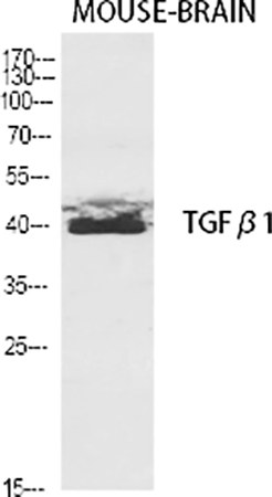
- Western Blot: Watch the Precursor vs. Mature vs. LAP
In reduced/denatured lysates, a clean anti‑TGF‑β1 should highlight:
• ~12.5 kDa band (mature dimer subunit) — your gold standard for "processed TGF‑β1."
• Sometimes ~45–50 kDa (non‑reduced dimer) or higher LAP‑containing bands if your lysis doesn't fully strip LAP or if you're probing non‑reduced gels.
Pro tip: If your band pattern looks like a smear across 35–55 kDa, ask whether you're seeing LAP cross‑reactivity or incomplete reduction. Run a positive control lysate (TGF‑β1–overexpressing cells or recombinant TGF‑β1) side‑by‑side to calibrate expectations.
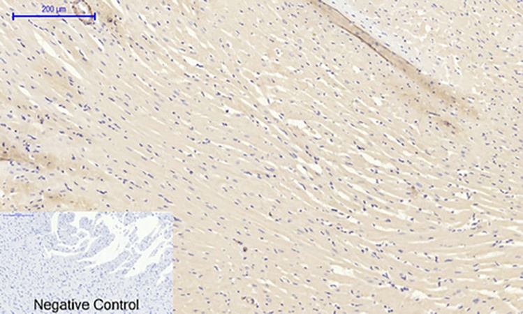
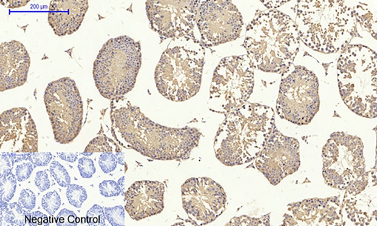
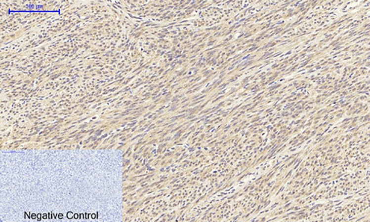
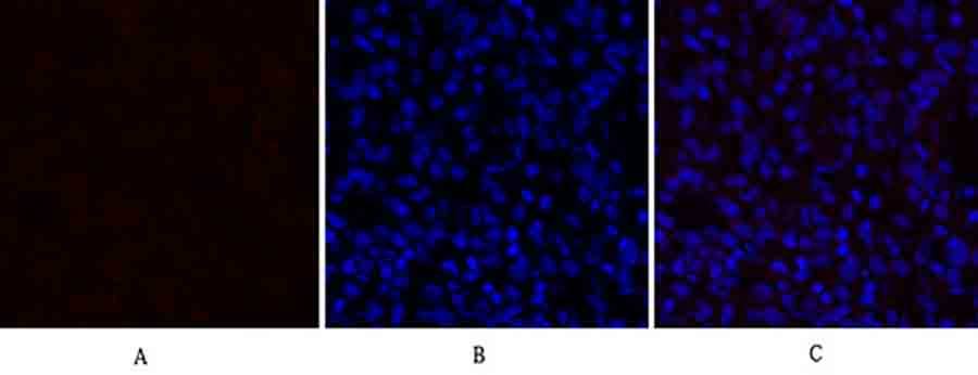
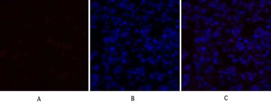
- IHC‑P / FFPE: It's All About Antigen Retrieval
TGF‑β1 sits in ECM as SLC/LTBP complexes, so formalin cross‑links can hide it. Most labs succeed with:
• pH 9.0 Tris/EDTA retrieval (heat‑mediated) for cytoplasmic/matrix TGF‑β1.
• Expect fine granular cytoplasmic + perimembrane/matrix staining; nuclear "TGF‑β1" staining is almost always background or a secondary artifact unless you have a very specific non‑canonical nuclear‑localization story and an orthogonal validation.
- Neutralization vs. Detection: Know Which Mode You're In
Because ABP52598 is an anti‑TGF‑β1 (not an anti‑LAP alone), it can be evaluated for functional blocking/neutralization in vitro—but you must confirm that experimentally (dose‑response on a TGF‑β‑responsive reporter) before treating it as a function‑blocking reagent. For ELISA, the cleaner play is often to coat/capture with an anti‑LAP or the supplied antibody (depending on whether you want total vs. active), then detect accordingly.
Where This Antibody Earns Its Keep in Real Research Programs
- Fibrosis pipelines (liver, lung, kidney, heart): Score TGF‑β1 upregulation in bleomycin/NASH/UUO models and patient biopsies with IHC/IF intensity + WB normalization—then tie it to p‑SMAD2/3 signaling and collagen I/α‑SMA readouts.
- Tumor‑microenvironment immunosuppression studies: Map TGF‑β1–rich zones adjacent to necrotic/hypoxic tumor fronts; combine with CD8/FoxP3/CD163 panels to argue "fibrotic, suppressive stroma."
- Wound healing & dermal scarring: Track TGF‑β1 appearance temporally post‑injury; validate with active‑TGF‑β1 ELISAs (acid‑activation vs. total) so you're not confusing latent stores with new burst.
- Immune‑cell polarization: If you're differentiating Th17 vs. iTreg and want a protein‑level anchor for why exogenous TGF‑β1 is skewing fate, ABP52598 gives you a direct probe for the added cytokine's presence/purity.
- Biomaterial & regenerative medicine: Scaffolds that bind/release TGF‑β1—use the antibody to confirm localized deposition or residual elution profile in vitro.
The Takeaway: In TGF‑β1 Biology, Your Antibody Is Your Interpretation
You can't separate "what TGF‑β1 is doing" from "how you're allowed to see it." The TGF‑β1 Polyclonal Antibody (ABP52598) from Abbkine is built for labs that need a versatile, affinity‑purified, multi‑application reagent—WB, IHC, IF, ELISA—without surrendering specificity for convenience. Treat it well (proper retrieval for IHC, good positive controls for WB, and a clear plan for latent vs. active), and it will repay you with the one thing this cytokine rarely gives away for free: an unambiguous signal.
Product Link: https://www.abbkine.com/product/tgf%ce%b21-polyclonal-antibody-abp52598/


