




TGF‑β1: The Master Regulator of Cellular Fate – How the EliKine™ Human TGF‑β1 ELISA Kit (KTE6030) Delivers Unmatched Sensitivity and Specificity in Cytokine Profiling
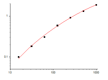
Transforming growth factor‑beta 1 (TGF‑β1) — the 25 kDa homodimeric cytokine that orchestrates cell proliferation, differentiation, apoptosis, immune regulation, and extracellular matrix remodeling — is not merely a signaling molecule; it is the pivotal switch that determines whether tissues heal or scar, tumors progress or regress, and immune responses are suppressed or activated, with circulating levels ranging from <10 pg/mL in healthy individuals to >500 pg/mL in fibrotic, metastatic, or autoimmune diseases . Yet, when you attempt to quantify TGF‑β1 in serum, plasma, cell culture supernatants, or tissue lysates using conventional ELISA kits, you encounter matrix interference from latent TGF‑β1 complexes, cross‑reactivity with TGF‑β2/β3 isoforms, and poor sensitivity that fails to capture physiologically relevant low‑abundance signals. The EliKine™ Human TGF‑β1 ELISA Kit (KTE6030) overcomes these limitations with a two‑site sandwich ELISA that detects active TGF‑β1 in human samples with 8 pg/mL sensitivity, a dynamic range of 15.6–1000 pg/mL, and zero cross‑reactivity with TGF‑β2, TGF‑β3, or other cytokines — enabling precise, high‑throughput TGF‑β1 quantification without acid activation, lengthy sample pretreatment, or signal amplification steps .
TGF‑β1 Is Not Just a Cytokine — It's the Bifunctional Orchestrator of Embryonic Development, Wound Healing, Immune Tolerance, and Disease Pathogenesis, with Dysregulation Driving Fibrosis, Cancer Metastasis, and Autoimmune Disorders
Transforming growth factor‑beta 1 (TGF‑β1), a member of the TGF‑β superfamily secreted as a latent complex (LTBP‑bound) that requires proteolytic or acidic activation, functions as a context‑dependent dual‑faced regulator . In normal physiology, TGF‑β1 maintains immune homeostasis by suppressing T‑cell proliferation, promoting regulatory T‑cell (Treg) differentiation, and inhibiting inflammatory cytokine production; it also drives wound healing by stimulating fibroblast migration, collagen deposition, and angiogenesis . However, in pathological states, sustained TGF‑β1 signaling induces epithelial‑to‑mesenchymal transition (EMT) in cancer cells, promoting metastasis; stimulates myofibroblast activation and excessive extracellular matrix (ECM) deposition, leading to fibrosis in lung, liver, and kidney; and contributes to autoimmune diseases like systemic sclerosis, rheumatoid arthritis, and lupus by disrupting immune tolerance . Clinically, serum TGF‑β1 levels correlate with tumor stage in breast, prostate, and colorectal cancers; fibrosis severity in idiopathic pulmonary fibrosis (IPF) and liver cirrhosis; and disease activity in systemic sclerosis . Yet, accurate TGF‑β1 measurement is confounded by the presence of latent TGF‑β1 (which constitutes >90% of circulating TGF‑β1), cross‑reactivity with TGF‑β2/β3 isoforms, and interference from serum proteins like α2‑macroglobulin, leading to either underestimation (if only active TGF‑β1 is detected) or overestimation (if latent complexes are inadvertently measured) .
Why Conventional TGF‑β1 ELISAs Fail in Complex Biological Matrices — And How the EliKine™ Kit (KTE6030) Uses a High‑Affinity, Isoform‑Specific Antibody Pair and Optimized Buffer System to Eliminate Cross‑Reactivity and Matrix Effects
The EliKine™ Human TGF‑β1 ELISA Kit (KTE6030) is a solid‑phase, two‑site sandwich enzyme‑linked immunosorbent assay (ELISA) that quantifies active TGF‑β1 in human serum, plasma, cell culture supernatants, and other biological fluids without requiring acid activation . The principle relies on a matched antibody pair: a monoclonal anti‑TGF‑β1 capture antibody is pre‑coated onto the microplate wells; after sample incubation, a biotin‑labeled detection antibody binds to a different epitope on TGF‑β1; then, streptavidin‑conjugated horseradish peroxidase (Streptavidin‑HRP) is added, followed by TMB substrate that produces a blue color proportional to TGF‑β1 concentration, which turns yellow upon acid stop and is measured at 450 nm . Unlike older kits that suffer from cross‑reactivity with TGF‑β2/β3 (which share ~70% sequence homology), interference from latent TGF‑β1‑binding proteins (LTBP, α2‑macroglobulin), and poor sensitivity (>20 pg/mL), this kit employs high‑affinity, isoform‑specific antibodies that recognize only active human TGF‑β1, optimized buffer systems that minimize non‑specific binding, and a Streptavidin‑HRP signal amplification system that enhances sensitivity to 8 pg/mL, enabling accurate TGF‑β1 measurement in a 3‑hour workflow .
Interference Source Effect on TGF‑β1 Measurement in Conventional ELISAs How KTE6030 Addresses It
Latent TGF‑β1 complexes Latent TGF‑β1 (bound to LTBP or α2‑macroglobulin) is not detected by most ELISAs, leading to underestimation of total TGF‑β1 The kit is designed to detect active TGF‑β1; for total TGF‑β1 measurement, samples can be acid‑activated (e.g., with 1 N HCl) and neutralized prior to assay, which releases active TGF‑β1 from latent complexes .
Cross‑reactivity with TGF‑β2/β3 Antibodies may bind TGF‑β2/β3, causing false‑high readings Uses isoform‑specific monoclonal antibodies that show no detectable cross‑reactivity with human TGF‑β2, TGF‑β3, or other cytokines (validated by Luminex xMAP technology) .
Matrix effects (serum, plasma) High‑abundance proteins (albumin, immunoglobulins) can cause non‑specific binding, increasing background Includes optimized blocking and wash buffers that reduce non‑specific binding; sample dilution (typically 1:2 to 1:10) further minimizes matrix interference .
Heterophilic antibodies (human anti‑mouse antibodies, HAMA) Endogenous antibodies in human samples may bridge capture and detection antibodies, causing false‑positive signals The antibody pair is derived from mouse; the kit includes blocking reagents to minimize HAMA interference; samples with suspected HAMA can be pre‑treated with heterophilic blocking tubes .
Low sensitivity Many commercial ELISAs have detection limits >20 pg/mL, missing physiologically relevant low‑level TGF‑β1 Achieves 8 pg/mL sensitivity through high‑affinity antibodies and Streptavidin‑HRP signal amplification, enabling detection of TGF‑β1 in normal serum (typically 10–50 pg/mL) .
Sample stability TGF‑β1 degrades rapidly at room temperature or after repeated freeze‑thaw cycles Recommends immediate freezing of samples at –80°C, avoiding repeated freeze‑thaw cycles, and adding protease inhibitors to preserve TGF‑β1 integrity .
Plate‑to‑plate variability Inconsistent coating or lot‑to‑lot antibody differences affect reproducibility Provides pre‑coated plates with QC‑tested consistency (CV <8% across batches) and lyophilized standards with defined potency .
The kit is validated for human serum, plasma (EDTA, heparin, citrate), cell culture supernatants (from fibroblasts, epithelial cells, immune cells), and other biological fluids (synovial fluid, bronchoalveolar lavage fluid, urine), with a dynamic range of 15.6–1000 pg/mL, covering both basal (10–50 pg/mL) and pathological (up to 1000 pg/mL) TGF‑β1 levels . It requires only 100 µL of sample per well (after dilution), enabling high‑throughput screening of clinical cohorts or limited‑volume samples (e.g., mouse serum, primary cell supernatants) .
What's in the Box (And the Three Critical Steps That Separate Accurate TGF‑β1 Quantification from Cross‑Reactivity and Matrix Artifacts)
Component Role in the Assay Handling & Storage
Pre‑coated microplate (96 wells) Mouse monoclonal anti‑human TGF‑β1 antibody immobilized on wells; captures TGF‑β1 from samples Store at 4°C in sealed bag with desiccant; stable until expiration date; bring to room temperature before use.
TGF‑β1 standard (lyophilized) Recombinant human TGF‑β1 protein (e.g., 1000 pg/mL) for generating standard curve Reconstitute with 1 mL of standard diluent to make stock solution; aliquot and store at –80°C; avoid repeated freeze‑thaw cycles.
Detection antibody (biotin‑labeled) Biotin‑conjugated mouse monoclonal anti‑human TGF‑β1 antibody; binds captured TGF‑β1 Store at 4°C; stable until expiration date; dilute as per protocol before use.
Streptavidin‑HRP conjugate Streptavidin‑horseradish peroxidase; binds biotin‑labeled detection antibody for signal amplification Store at 4°C; stable until expiration date; dilute as per protocol before use.
TMB substrate solution 3,3',5,5'‑tetramethylbenzidine (TMB); HRP catalyzes oxidation to blue product Store at 4°C protected from light; bring to room temperature before use; color develops within 15–30 minutes.
Stop solution (acid) 1 N sulfuric acid or similar; stops HRP reaction and turns blue to yellow Store at room temperature; corrosive—handle with care.
Wash buffer (20× concentrate) PBS‑Tween 20 or similar; removes unbound reagents Dilute to 1× with distilled water before use; store diluted buffer at 4°C for up to 1 week.
Assay diluent Buffer for diluting samples and standards; reduces matrix interference Store at 4°C; bring to room temperature before use.
Plate sealer Adhesive film to cover plate during incubations Store at room temperature.
Key procedural steps that dictate accuracy:
Step Purpose Common Pitfalls & Solutions
- Sample collection & preparation Collect serum/plasma without hemolysis; prepare cell supernatants without TGF‑β1 degradation Collect blood in serum separator tubes or EDTA/heparin tubes; separate serum/plasma within 2 hours; store at –80°C; for cell supernatants, centrifuge to remove debris and store at –80°C; avoid repeated freeze‑thaw cycles.
- Standard curve preparation Generate a 7‑point standard curve (e.g., 1000, 500, 250, 125, 62.5, 31.25, 15.6 pg/mL) Reconstitute standard with 1 mL of assay diluent; perform serial 1:2 dilutions; include a zero standard (assay diluent only); prepare fresh for each assay.
- Plate incubation Allow TGF‑β1 in samples to bind to capture antibody Add 100 µL of standard or diluted sample to wells; cover with plate sealer; incubate at 37°C for 90 minutes (or room temperature for 2 hours).
- Detection antibody incubation Bind biotin‑labeled detection antibody to captured TGF‑β1 After washing, add 100 µL of detection antibody; cover; incubate at 37°C for 60 minutes (or room temperature for 90 minutes).
- Streptavidin‑HRP incubation Bind Streptavidin‑HRP to biotin‑labeled antibody After washing, add 100 µL of Streptavidin‑HRP; cover; incubate at 37°C for 30 minutes (or room temperature for 45 minutes).
- TMB substrate & stop Develop color and stop reaction After washing, add 100 µL of TMB substrate; incubate in dark at room temperature for 15–30 minutes; add 50 µL of stop solution; read at 450 nm within 30 minutes.
- Data analysis Convert absorbance to concentration Plot standard curve (log TGF‑β1 concentration vs. absorbance); fit 4‑parameter logistic (4PL) curve; interpolate sample concentrations; multiply by dilution factor.
- Normalization Express results per volume, cell count, or protein content For serum/plasma: pg/mL; for cell supernatants: pg/mL or pg/10⁶ cells; for tissue lysates: pg/mg protein.
The 3‑Hour Protocol That Turns KTE6030 into a Routine Tool for Immunology, Oncology, and Fibrosis Research
- Sample preparation
• For serum/plasma: Thaw frozen samples on ice; centrifuge at 10,000 × g for 10 min to remove any precipitate; dilute 1:2 to 1:10 with assay diluent to bring TGF‑β1 concentration within range (15.6–1000 pg/mL).• For cell culture supernatants: Collect supernatant from cells stimulated with TGF‑β1 inducers (e.g., TGF‑β1, LPS, TNF‑α); centrifuge at 2000 × g for 10 min to remove cells/debris; dilute as needed.• For tissue homogenates: Homogenize 50–100 mg of tissue in 1 mL of lysis buffer (e.g., RIPA with protease inhibitors); centrifuge at 12,000 × g for 15 min at 4°C; collect supernatant; dilute as needed.• Optional acid activation for total TGF‑β1: Add 10 µL of 1 N HCl to 100 µL of sample, incubate at room temperature for 10 minutes, then neutralize with 10 µL of 1.2 N NaOH/0.5 M HEPES; proceed with assay. - Reagent preparation
• Bring all reagents to room temperature (30 minutes before assay).• Prepare 1× wash buffer by diluting 20× concentrate with distilled water.• Prepare standard curve by serial dilution of reconstituted TGF‑β1 standard (1000 pg/mL) in assay diluent. - Assay procedure (96‑well plate format)
• Add 100 µL of standard or diluted sample to appropriate wells.• Cover with plate sealer; incubate at 37°C for 90 minutes.• Wash plate 4 times with 300 µL/well of 1× wash buffer.• Add 100 µL of detection antibody to each well; cover; incubate at 37°C for 60 minutes.• Wash plate 4 times.• Add 100 µL of Streptavidin‑HRP to each well; cover; incubate at 37°C for 30 minutes.• Wash plate 4 times.• Add 100 µL of TMB substrate; incubate in dark at room temperature for 15–30 minutes (until blue color develops).• Add 50 µL of stop solution; read absorbance at 450 nm within 30 minutes. - Calculation
• Subtract blank absorbance (zero standard) from all readings.• Plot standard curve using 4‑parameter logistic (4PL) fit (log concentration vs. absorbance).• Interpolate sample concentrations from curve.• Multiply by dilution factor to obtain original concentration.
What Actually Changes When You Switch from Conventional TGF‑β1 ELISAs to the EliKine™ Kit
① Your sensitivity improves from >20 pg/mL to 8 pg/mL, allowing you to detect basal TGF‑β1 levels in normal serum and subtle changes in early‑stage fibrosis or minimal residual disease.
Many commercial ELISAs have detection limits of 20–50 pg/mL, missing the 10–50 pg/mL range seen in healthy individuals; the EliKine™ kit's high‑affinity antibodies and Streptavidin‑HRP amplification lower the limit to 8 pg/mL, enabling detection of physiologically relevant low‑abundance TGF‑β1 .
② You eliminate cross‑reactivity with TGF‑β2 and TGF‑β3, because the kit's isoform‑specific monoclonal antibodies show zero detectable binding to other TGF‑β isoforms, giving you true TGF‑β1‑specific signals.
TGF‑β2 and TGF‑β3 share high sequence homology and can cross‑react in polyclonal‑based ELISAs; the monoclonal antibody pair in KTE6030 is rigorously validated to show no cross‑reactivity, ensuring specific quantification of TGF‑β1 .
③ You can process serum, plasma, and cell culture supernatants without matrix interference headaches, thanks to optimized buffers that reduce non‑specific binding and a pre‑coated plate format that minimizes inter‑assay variability.
Serum proteins (albumin, immunoglobulins) can cause high background; the kit's blocking buffers and wash steps effectively reduce non‑specific binding, while pre‑coated plates ensure consistent coating across wells and batches.
④ Your data become reproducible across operators and batches because the kit standardizes antibody lots, plate coating, and reagent stability, with inter‑assay CV <8%.
Inter‑lab variation in TGF‑β1 ELISAs often stems from inconsistent antibody affinity, plate coating efficiency, and TMB lot differences; the EliKine™ kit provides QC‑tested, pre‑coated plates and lyophilized standards with batch‑to‑batch consistency .
Where KTE6030 Earns Its Place in the Lab's Routine Immunology, Oncology, and Fibrosis Research Panels
Application Why Precise TGF‑β1 Quantification Is Critical
Cancer biology & metastasis Measure TGF‑β1 in tumor tissue, serum, or cell supernatants to assess EMT induction, angiogenesis, and immune evasion in breast, prostate, colorectal, and pancreatic cancers .
Fibrosis research (lung, liver, kidney) Quantify TGF‑β1 in bronchoalveolar lavage fluid (BALF), liver homogenates, or urine to evaluate fibrosis progression and response to anti‑fibrotic therapies (pirfenidone, nintedanib) .
Autoimmune disease monitoring Monitor serum TGF‑β1 in systemic sclerosis, rheumatoid arthritis, lupus to correlate with disease activity, skin thickness, and organ involvement .
Wound healing & tissue repair Assess TGF‑β1 in wound fluid, fibroblast supernatants, or skin biopsies to study collagen synthesis, myofibroblast differentiation, and scar formation .
Immunology & T‑cell regulation Detect TGF‑β1 secreted by regulatory T cells (Tregs), dendritic cells, or macrophages to investigate immune suppression, tolerance, and inflammation .
Stem cell & developmental biology Measure TGF‑β1 in embryonic stem cell cultures, organoids, or developmental models to understand lineage specification, morphogenesis, and differentiation .
Drug screening for TGF‑β pathway inhibitors Screen for TGF‑β receptor kinase inhibitors (galunisertib, vactosertib), neutralizing antibodies (fresolimumab), or siRNA using TGF‑β1 as a pharmacodynamic biomarker.
Clinical research & biomarker discovery Analyze TGF‑β1 in large cohort studies as a prognostic marker for cancer recurrence, fibrosis progression, or autoimmune flare‑ups (research use only).
Cardiovascular research Evaluate TGF‑β1 in cardiac fibroblasts, atherosclerotic plaques, or myocardial infarction models to study cardiac remodeling, fibrosis, and heart failure .
Neuroscience & neuroinflammation Quantify TGF‑β1 in cerebrospinal fluid (CSF), microglial supernatants, or brain homogenates to investigate neuroprotection, glial activation, and neurodegenerative diseases .
A Drop‑In Methods Paragraph
TGF‑β1 concentration was determined using the EliKine™ Human TGF‑β1 ELISA Kit (KTE6030, Abbkine) according to the manufacturer's protocol. Briefly, 100 µL of serum (diluted 1:5 with assay diluent) or recombinant TGF‑β1 standard (0–1000 pg/mL) was added to a pre‑coated 96‑well plate. After incubation at 37°C for 90 minutes and washing, 100 µL of biotin‑labeled detection antibody was added and incubated at 37°C for 60 minutes. Following another wash, 100 µL of Streptavidin‑HRP was added and incubated at 37°C for 30 minutes. After a final wash, 100 µL of TMB substrate was added and incubated in the dark for 20 minutes. The reaction was stopped with 50 µL of stop solution, and absorbance was measured at 450 nm using a microplate reader (Bio‑Rad iMark). TGF‑β1 concentration was calculated by interpolating from a 4‑parameter logistic standard curve. All samples were assayed in duplicate, and the intra‑assay coefficient of variation (CV) was <10%.
The Bench Rules That Keep Your TGF‑β1 Measurements Accurate and Reproducible
Rule Why It Matters
🧪 Always run a fresh standard curve with each assay (7 points in duplicate) TGF‑β1 standards degrade over time; never reuse curves from previous runs.
⏱️ Read absorbance within 30 minutes after adding stop solution The yellow color may fade or precipitate over time, leading to inaccurate readings.
🧊 Store samples at –80°C if not assayed immediately; avoid repeated freeze‑thaw cycles TGF‑β1 is prone to degradation and adsorption to tube walls; aliquot samples to minimize freeze‑thaw.
⚗️ Include a sample blank (assay diluent only) and a negative control (unstimulated cell supernatant) Correct for background absorbance and non‑specific binding.
🔬 For total TGF‑β1 measurement, perform acid activation (1 N HCl) followed by neutralization Latent TGF‑β1 constitutes >90% of circulating TGF‑β1; acid activation releases active TGF‑β1 for detection.
🚫 Avoid hemolyzed or lipemic samples Hemolysis releases erythrocyte proteases that degrade TGF‑β1; lipemia can cause non‑specific binding.
🧼 Use low‑binding tubes and pipette tips TGF‑β1 adsorbs to plastic surfaces; use siliconized or low‑binding consumables to minimize loss.
📊 Include a quality‑control sample (pooled human serum or commercial control) in each run Monitor inter‑assay precision and detect systematic drift.
🌡️ Bring all reagents to room temperature before use Temperature variations affect antibody binding and enzymatic reaction kinetics.
🧪 Perform sample dilution to ensure readings fall within the standard curve range Samples with TGF‑β1 >1000 pg/mL may require further dilution to avoid hook effect.
Explore the EliKine™ Human TGF‑β1 ELISA Kit (KTE6030) full specifications, protocol, and ordering options here:
🔗 https://www.abbkine.com/product/elikine-human-tgf-%ce%b21-elisa-kit-kte6030/
(For research use only. Not for human or clinical diagnostic use. Store at 4°C protected from light; bring reagents to room temperature before use; avoid freeze‑thaw cycles; always include a standard curve; read absorbance within 30 minutes after stopping.)





