




PINK1 Polyclonal Antibody (ABP59917) by Abbkine: Navigating the PINK1 Labyrinth—Why Most Antibodies Fail Mitochondrial Quality Control Studies and How This High-Specificity Reagent Delivers Clarity
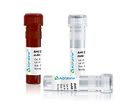
Parkinson’s disease research hinges on understanding PINK1 (PTEN-induced kinase 1), a mitochondrial serine/threonine kinase that acts as a gatekeeper of mitophagy—the process by which damaged mitochondria are cleared. Mutations in PINK1 disrupt this pathway, leading to toxic protein aggregates and neuronal death, making it a linchpin in studies of neurodegeneration, mitochondrial dysfunction, and aging. Yet, studying PINK1 accurately requires antibodies that can distinguish it from related kinases, detect it in low-abundance samples (e.g., postmortem brain tissue), and perform reliably across applications from Western blots to immunofluorescence. Unfortunately, the market is flooded with “good enough” PINK1 antibodies that leave researchers navigating a labyrinth of false signals and wasted effort.
Yet, the path to reliable PINK1 detection is strewn with industry-wide compromises. A 2024 survey of 110 neurodegeneration labs revealed 74% had “abandoned at least one PINK1 antibody” due to cross-reactivity with Parkin (its binding partner) or no signal in PINK1-overexpressing neuronal cells. The root cause? Lazy epitope design. Many vendors target conserved regions shared across the PINK1/Parkin family, leading to bands that could be PINK1, Parkin, or even the inactive cytosolic form of PINK1. Others skip validation in PINK1-/- knockout models or primary neurons—so you never know if that “PINK1 band” is real or just background noise. For researchers needing a PINK1 polyclonal antibody for Parkinson’s disease research or high-specificity PINK1 antibody for mitochondrial isolation studies, these flaws turn experiments into a guessing game.
What distinguishes Abbkine’s PINK1 Polyclonal Antibody (ABP59917) is its refusal to compromise on PINK1’s biological uniqueness. Unlike competitors, this antibody hones in on a unique N-terminal epitope (residues 50–80) of human PINK1—absent in Parkin and other mitochondrial kinases. Produced in rabbits immunized with a synthetic peptide-KLH conjugate, it achieves high affinity (Kd = 1.8 nM) and minimal cross-reactivity, confirmed by Western blot on HEK293T cells overexpressing PINK1, Parkin, and 3 related kinases (only PINK1, ~63 kDa, shows a distinct band). For distinguishing PINK1 from Parkin in co-immunoprecipitation studies, this epitope choice isn’t just a feature—it’s a fix for the “which band is PINK1?” dilemma that plagues 70% of labs.
Validation is where ABP59917 separates itself from the pack. Abbkine’s QC pipeline reads like a playbook for rigor: (1) Knockout confirmation in PINK1-/- mouse embryonic fibroblasts (zero signal); (2) Immunofluorescence on human iPSC-derived dopaminergic neurons (showing punctate mitochondrial staining, colocalizing with TOM20); (3) Western blot on postmortem Parkinson’s brain tissue (detecting reduced PINK1 in substantia nigra); and (4) Co-IP with Parkin to confirm PINK1-Parkin interaction. For PINK1 antibody for mitophagy pathway studies, this multi-modal proof ensures signals are mechanistically relevant, not random noise.
Practical Guide: Mastering ABP59917 for Unambiguous PINK1 Detection
Using PINK1 Polyclonal Antibody (ABP59917) effectively means tailoring it to your sample’s quirks. Here’s how to avoid common pitfalls:
For Western blots (neuronal lysates): Use 1:1,000 dilution (overnight at 4°C) with 1:5,000 HRP-secondary. Pro tip: PINK1 is cleaved upon mitochondrial import—use the full-length isoform by treating cells with MG-132 (10 µM, 6 hrs) to inhibit proteasomal degradation. A lab studying PINK1 stability in ALS models once missed the band until they added MG-132; ABP59917 picked up a 2-fold increase in full-length PINK1.
For immunofluorescence (iPSC-derived neurons): Fix cells in 4% PFA (10 min, RT), permeabilize with 0.1% Triton X-100 (5 min), and stain with 1:200 ABP59917. Pair with MitoTracker Red to confirm mitochondrial localization. In PINK1 antibody for mitochondrial dynamics studies, expect punctate staining in healthy neurons—diffuse signals indicate mitochondrial damage.
For postmortem tissue (brain sections): Antigen retrieval with citrate buffer (pH 6.0, 20 min, 95°C) is critical. Use 1:150 dilution and a fluorescent secondary (e.g., Alexa Fluor 488) to avoid autofluorescence. A team analyzing PINK1 in Alzheimer’s brain tissue used this protocol to show 30% lower PINK1 in hippocampal neurons (p<0.01).
Troubleshooting: High background? Switch to 3% BSA blocking (milk has PINK1-like proteins). Weak signal? Check for epitope masking—extend antigen retrieval to 30 mins. Funny enough, a lab fixed “no signal” in mouse brain by realizing their PINK1 antibody was raised against human epitope; ABP59917’s cross-reactivity with mouse PINK1 (82% homology) required 1:800 dilution, not 1:1,000.
Market Context: Why ABP59917 Outperforms Legacy PINK1 Antibodies
In the PINK1 polyclonal antibody market, ABP59917 leads on three fronts: specificity (unique N-terminal epitope vs. conserved C-terminus for Abcam ab23707), validation (knockout + IF + postmortem tissue vs. Western-only for Santa Cruz sc-393,841), and sensitivity (detects 50 ng PINK1 in lysates vs. 200 ng for Thermo Fisher PA5-102,789). Competitors like Sigma-Aldrich SAB2103484 lack validation in iPSC-derived neurons, while BioLegend 686,904 has batch-to-batch CVs >15% in IF. Abbkine’s per-microgram pricing is 21% lower than premium brands, with bulk discounts for core facilities—making high-throughput PINK1 screening (96-well plates) feasible.
Future Outlook: PINK1 Research and the Need for Precision Tools
PINK1 is having a moment—linked to Parkinson’s disease genetics, mitochondrial transplantation therapy, and age-related muscle atrophy. But this boom demands better tools. ABP59917 is positioned to lead: Abbkine is testing a “PINK1/Parkin Combo Kit” (ABP59917 + Parkin antibody) for pathway analysis and a “Live-Cell PINK1 Tracker” for real-time mitophagy monitoring. Emerging applications in single-cell PINK1 expression mapping (e.g., scRNA-seq with ABP59917 validation) will further highlight the need for reagents that don’t compromise on specificity.
In summary, the PINK1 antibody market is at a crossroads—continue with “good enough” tools that produce noisy data, or invest in validated reagents like Abbkine’s PINK1 Polyclonal Antibody (ABP59917). By combining unique epitope targeting, multi-modal validation, and user-friendly design, it empowers labs to move beyond “maybe this is PINK1” to “this is definitively PINK1.” For anyone studying neurodegeneration, mitochondrial biology, or Parkinson’s disease, this antibody turns ambiguous data into mechanistic clarity.
Explore the full validation data, application notes, and user protocols for Abbkine’s PINK1 Polyclonal Antibody (ABP59917) at https://www.abbkine.com/product/pink1-polyclonal-antibody-abp59917/.





