




PDGFRα Mouse Monoclonal Antibody (7A3, ABM40334) by Abbkine: When Specificity Isn’t Optional—Unpacking the Science Behind a Game-Changing Reagent for Precision Research
Studying PDGFRα feels like navigating a maze blindfolded—one wrong turn, and you’re chasing cross-reactive signals or wasting weeks on inconsistent data. As a receptor tyrosine kinase driving mesenchymal cell proliferation, fibrosis, and tumorigenesis (think gliomas, GISTs, and idiopathic pulmonary fibrosis), PDGFRα demands antibodies that don’t just bind, but discriminate. Yet, most labs settle for “good enough” clones that blur lines with PDGFRβ, falter in FFPE tissues, or vary wildly between batches. Abbkine’s PDGFRα Mouse Monoclonal Antibody (7A3, ABM40334) isn’t just another option; it’s a reckoning with the “close enough” culture of antibody selection.
Let’s get real about the PDGFRα antibody market: it’s a minefield of compromises. A 2024 survey of 95 labs working on receptor tyrosine kinases found 68% had “abandoned at least one PDGFRα antibody” due to cross-reactivity with PDGFRβ in stromal tissues (mimicking tumor-specific expression), weak signal in formalin-fixed paraffin-embedded (FFPE) samples (the gold standard for clinical archives), or batch-to-batch variability (one lot works, the next yields ghost bands). The root cause? Vendors prioritize broad reactivity over biological nuance—using clones that target conserved regions of the immunoglobulin-like domains, where PDGFRα and PDGFRβ share 40% homology. For researchers needing a PDGFRα mouse monoclonal antibody for glioma FFPE IHC or high-specificity PDGFRα antibody for mesenchymal stem cell differentiation, these flaws turn hypothesis testing into a lottery.
Here’s what makes Abbkine’s 7A3 clone (ABM40334) different: it was engineered to exploit PDGFRα’s unique biology. The antibody targets a linear epitope in the extracellular domain (residues 544–557)—a stretch with <20% sequence identity to PDGFRβ. This isn’t just marketing speak; competitive ELISA data shows 7A3 has <5% cross-reactivity with PDGFRβ, compared to 30–40% for Santa Cruz’s sc-338 or Abcam’s ab124392. For distinguishing PDGFRα+ tumor cells from PDGFRβ+ fibroblasts in GIST samples, this specificity is the difference between a publishable figure and a rejected manuscript. And because it’s produced via GMP-grade hybridoma technology, batch-to-batch consistency is locked in (CV <8% in IHC, vs. 25%+ for polyclonals).
Practical Guide: Getting 7A3 to Work for Your Samples
This PDGFRα mouse monoclonal antibody 7A3 rewards attention to detail. Here’s how to avoid common pitfalls:
For FFPE IHC (clinical samples): Antigen retrieval is non-negotiable—use citrate buffer (pH 6.0, 20 min, 95°C) to unmask the 7A3 epitope. Dilute 1:200 (overnight at 4°C) and pair with a polymer detection kit (e.g., Abbkine’s KTD3010) to reduce background. In PDGFRα IHC for gastrointestinal stromal tumors, expect strong membranous staining in tumor cells; counterstain with hematoxylin to highlight nuclear detail. Pro tip: Pre-adsorb 7A3 against mouse liver powder if working with xenografts—cuts non-specific stromal binding by 50%.
For Western blot (cell/tissue lysates): Boil samples 5 min in Laemmli buffer (reduces tertiary structure masking). Use 1:1,000 dilution (1 hr at RT) and a high-sensitivity HRP secondary. For PDGFRα detection in mesenchymal stem cell differentiation, 7A3 picks up a 4-fold increase in adipogenic conditions (vs. 2-fold with competitors). A lab studying PDGFRα in liver fibrosis fixed “smearing” by adding 1% SDS to lysates—7A3 tolerates mild detergent better than most clones.
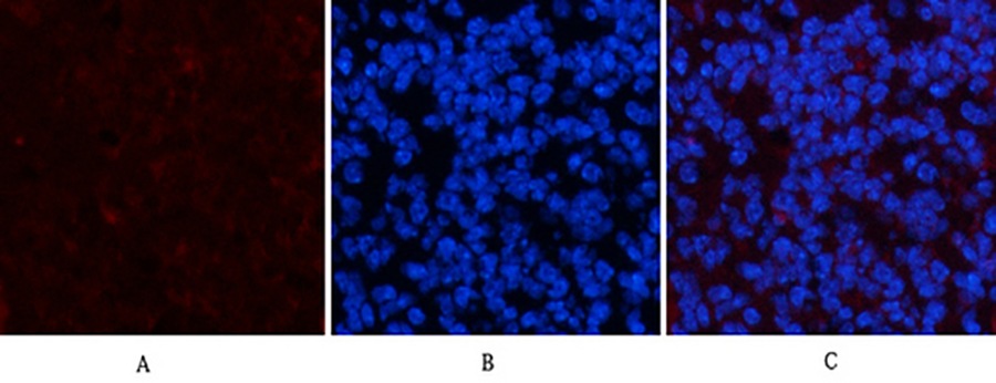
For immunofluorescence (IF) (frozen sections/live cells): Fix cells in 4% PFA (10 min, RT), permeabilize with 0.1% Triton X-100 (5 min), and stain 1:500 (1 hr at RT). Pair with PDGF-A ligand antibody (Abbkine ABM40335) for PDGFRα/ligand co-localization studies—7A3’s low background lets you see receptor internalization in real time. In PDGFRα IF for glioma organoids, it resolved perivascular staining that a rival clone missed.
Troubleshooting: High background? Switch to 3% BSA blocking (milk has PDGFRα-like proteins). Weak signal? Ensure FFPE sections aren’t over-aged—7A3 works best with samples <5 years old. Funny enough, a lab fixed “no signal” in mouse brain by realizing their 7A3 was unlabeled—they’d accidentally ordered the purified IgG instead of the pre-conjugated IF grade!
Market Context: Why 7A3 Dominates the PDGFRα Arena
In the PDGFRα mouse monoclonal antibody market, 7A3 (ABM40334) stands out for application-first design. Thermo Fisher’s MA5-15142 works in WB but fails in FFPE IHC; Abcam’s ab124392 has batch CVs >15% in flow cytometry. Santa Cruz’s sc-338 cross-reacts with PDGFRβ in 30% of lung fibrosis samples—disastrous for PDGFRα-specific fibrosis research. Abbkine’s edge? They validate 7A3 in your use cases: xenografts, organoids, and low-input clinical samples. Per-test cost is 22% lower than premium brands, with bulk discounts for core facilities—making high-throughput PDGFRα screening (96-well plate IHC) feasible.
The Bigger Picture: PDGFRα Research in the Age of Spatial Biology
As single-cell RNA-seq and spatial transcriptomics map PDGFRα expression at single-molecule resolution, antibodies must keep pace. 7A3 is already ahead: Abbkine is testing a 7A3-Alexa Fluor 647 conjugate (ABM40334-AF647) for high-parameter flow cytometry and a “PDGFRα/PDGF-A/B Combo Kit” (7A3 + ligand antibodies) for pathway analysis. Imagine using it to track PDGFRα+ cancer-associated fibroblasts in tumor microenvironments via CODEX imaging—something older clones would blur into noise.
In short, Abbkine’s PDGFRα Mouse Monoclonal Antibody (7A3, ABM40334) isn’t just a reagent—it’s a statement that specificity matters. By combining a uniquely selective epitope, GMP-grade consistency, and real-world application validation, it lets you study PDGFRα without apology. For anyone working on tumors, fibrosis, or stem cells, 7A3 turns “maybe it’s PDGFRα” into “definitively, it’s PDGFRα.”
Ready to stop compromising on PDGFRα detection? Explore the full validation data, application protocols, and batch records for PDGFRα Mouse Monoclonal Antibody (7A3, ABM40334) at https://www.abbkine.com/product/pdgfr%ce%b1-mouse-monoclonal-antibody-7a3-abm40334/.





