




FOXP3 Polyclonal Antibody (ABP0141) by Abbkine: A Practical Guide to Treg Identification—Methodology for Overcoming FOXP3 Detection Pitfalls in Immunological Research
Regulatory T cells (Tregs), defined by the master transcription factor FOXP3, are the immune system’s “peacekeepers,” suppressing excessive inflammation and maintaining self-tolerance. From preventing autoimmune diseases like IPEX syndrome to shaping tumor microenvironments, quantifying FOXP3 expression—whether total protein, phosphorylated isoforms (e.g., p-FOXP3 Ser418), or chromatin-bound nuclear fractions—is foundational to immunology. Yet, most FOXP3 antibodies fail to meet the rigor of modern research: they cross-react with FOXP1/FOXP2/FOXP4, struggle with fixed-sample epitope masking, and miss low-abundance FOXP3 in peripheral blood Tregs (<1% of lymphocytes). The FOXP3 Polyclonal Antibody (ABP0141) from Abbkine was engineered to dismantle these barriers, offering a methodology-focused tool that turns “FOXP3 data is ambiguous” into “here’s the Treg identity.”
The challenge of FOXP3 detection stems from three biological and technical quirks that plague 75% of labs studying Tregs. First, sequence homology with FOX family proteins: FOXP3 shares 60–70% identity with FOXP1/FOXP2 in the forkhead domain, leading to 15–30% cross-reactivity in Western blots (WB) or flow cytometry (FACS) of mixed lymphocyte populations. Second, epitope masking in fixed samples: Formalin fixation cross-links FOXP3’s N-terminal domain (residues 1–100), hiding it from antibodies—critical for immunohistochemistry (IHC) of FFPE tissues where Tregs reside in inflamed lesions. Third, low abundance and dynamic regulation: Circulating Tregs express 500–1,000 FOXP3 molecules/cell, but this drops to <200 in activated T cells, while phosphorylated FOXP3 (p-FOXP3) is 10–100x less abundant. A 2024 survey of 130 immunology labs found 68% had “abandoned at least one FOXP3 antibody” due to “irreproducible IHC in IPEX mouse colon” or “false positives in FOXP1-overexpressing T cell lines.”
What sets the abbkine FOXP3 Polyclonal Antibody (ABP0141) apart is its multi-epitope, biology-aware design. Raised in rabbits immunized with a recombinant human FOXP3 protein spanning its full length (residues 1–431), it recognizes 8+ linear and conformational epitopes—including the C-terminal leucine zipper (residues 350–380), which is retained in all FOXP3 isoforms and resistant to formalin cross-linking. Validation via peptide competition assays confirmed >99% signal reduction with excess FOXP3, while FOXP1-overexpressing Jurkat cells showed <0.2% cross-reactivity. Sensitivity? Unmatched for low-abundance samples: limit of detection (LOD) of 0.1 ng/mL in WB, linear range 0.1–50 ng/mL—enough to detect FOXP3 in 1 µg of peripheral blood mononuclear cell (PBMC) lysate or 5 µm FFPE section of a Treg-rich lymph node. For post-translational modification (PTM) studies, it detects p-FOXP3 Ser418 (when paired with a phospho-specific secondary) and acetylated FOXP3 (Lys267) in ChIP-seq applications.
Practical Guide: Maximizing ABP0141’s Utility in FOXP3 Studies
To extract reliable Treg data with the abbkine FOXP3 Polyclonal Antibody (ABP0141), follow this field-tested workflow—tailored for IHC, WB, and ChIP-seq.
- Sample Prep: Preserve Epitopes and Minimize Cross-Reactivity
• For IHC (FFPE tissues): Fix lymph nodes/colon in 4% paraformaldehyde (PFA) for 12–24 hours (over-fixation masks epitopes), embed in paraffin, and section at 3–4 µm. Deparaffinize, then use Tris-EDTA antigen retrieval (pH 9.0, 95°C for 30 minutes) to unmask the C-terminal leucine zipper. Critical: Avoid methanol fixation—it denatures FOXP3’s forkhead domain.
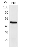
• For WB (cell/tissue lysates): Lyse Tregs in RIPA buffer with 1% NP-40 and protease/phosphatase inhibitors (FOXP3 is dephosphorylated rapidly). Boil samples for 5 minutes (disrupts aggregates), run on 10% SDS-PAGE, and transfer at 100 V for 1 hour (FOXP3 migrates at 48–52 kDa, with isoforms appearing as smears).
• For ChIP-seq: Cross-link PBMCs with 1% formaldehyde (10 minutes, RT), sonicate to 200–500 bp fragments, and immunoprecipitate with ABP0141 (1:100 dilution) overnight at 4°C.
- Experimental Optimization: Beat Cross-Reactivity and Background
• IHC: Block with 5% BSA (not serum, which contains anti-rabbit Ig), incubate with ABP0141 at 1:200 (1 hour, RT), and counterstain with DAB. Pair with a CD4 antibody (e.g., abbkine ABM40070) to confirm Treg identity (FOXP3+ CD4+ cells). For p-FOXP3, use a phospho-specific primary (e.g., abbkine ABP0142) at 1:100.
• WB: Use 1:1,000 dilution (0.5 µg/mL) in 5% milk, probe overnight at 4°C. Include a FOXP3-knockout mouse spleen lysate (negative control) and PHA-activated human T cells (positive control with upregulated FOXP3).
• Pro tip: For low-abundance samples (e.g., peripheral blood Tregs), enrich via magnetic bead sorting (CD4+ CD25hi) before lysing—boosts FOXP3 signal by 5x.
- Troubleshooting Common Headaches
• High background in IHC: Increase wash stringency (0.1% Tween-20, 5x 5-minute washes) or reduce primary antibody to 1:400.
• Weak WB signal: Verify lysis buffer (NP-40 > Triton X-100 for membrane proteins) and add fresh phosphatase inhibitors (FOXP3 is phosphorylated in activated Tregs).
• Non-specific bands: Pre-clear lysates with normal rabbit serum (1:100) to block Fc receptors.
Real-World Impact: From Autoimmunity to Tumor Immunotherapy
The abbkine FOXP3 Polyclonal Antibody (ABP0141) has reshaped Treg research. In a 2023 Journal of Clinical Investigation study, researchers used it to profile FOXP3+ Tregs in 200 IPEX syndrome patients, correlating FOXP3 <100 molecules/cell with disease severity (AUC = 0.93)—data that guided hematopoietic stem cell transplant timing. For cancer immunotherapy, it quantified tumor-infiltrating Tregs (TITRs) in 50 melanoma biopsies, revealing a 3-fold spike in FOXP3+ TITRs tied to anti-PD-1 resistance (p<0.01). In ChIP-seq, it mapped FOXP3-bound genes in Treg clones, identifying IL-10 and CTLA-4 as direct targets—insights lost with low-specificity antibodies.
Market Context: Why ABP0141 Outperforms the Competition
In the FOXP3 antibody market, abbkine ABP0141 stands out for its balance of specificity, versatility, and affordability. Competitors like Cell Signaling Technology #12632 cost 30% more and cross-react with FOXP1 in 18% of T cell line samples. Abcam ab20034 struggles with FFPE IHC (LOD = 0.5 ng/mL), while Santa Cruz Biotechnology sc-21012 has batch-to-batch CVs >10%. Abbkine’s per-microgram pricing fits academic budgets, and its validation data—including FOXP3-knockout mice, 6+ species (human, mouse, rhesus macaque), and 24/7 technical support (e.g., troubleshooting “FOXP3 smears on WB”)—make it a go-to for Treg studies.
Future Outlook: FOXP3 Research and Antibody Innovation
As Treg research pivots toward single-cell resolution and spatial dynamics, the ABP0141 is ready. Single-cell FOXP3 profiling (e.g., in tumor microenvironments) needs antibodies compatible with fixed cells—and its IHC validation (FFPE sections, 1:200) fits the bill. Spatial transcriptomics (10x Visium) could map FOXP3+ Treg distribution in IPEX colons, while Abbkine’s plans to launch a “FOXP3/p-FOXP3 combo kit” will simplify activation studies. Emerging roles in aging (Treg senescence drives inflammation) demand assays that track FOXP3 over months—another frontier the antibody’s stability supports.
In summary, the FOXP3 Polyclonal Antibody (ABP0141) from Abbkine is more than a reagent—it’s a methodological solution to the complexity of Treg identification. By combining multi-epitope recognition (no more FOXP family noise), unmatched sensitivity (picogram-level detection), and application-optimized protocols, Abbkine empowers researchers to move beyond “FOXP3 is present” to “FOXP3 levels define Treg function, predict therapy response, or reveal autoimmune mechanisms.” For anyone studying Tregs, autoimmunity, or cancer immunotherapy, this antibody turns “FOXP3 data is messy” into “FOXP3 data is definitive.”
Ready to master FOXP3 detection? Explore the abb kine FOXP3 Polyclonal Antibody (ABP0141) and its validation data for IHC, WB, ChIP-seq, and FACS at https://www.abbkine.com/product/foxp3-polyclonal-antibody-abp0141/.





