


The Original Danger Signal: Why HMGB1 (HMG-1) Remains the Most Powerful — and Dangerous — Protein in Your Lysate
If your lab works on inflammation, trauma, sepsis, autoimmunity, or tumor microenvironments and you're still treating HMGB1 as a "housekeeping control that accidentally ended up in your blot," you are sitting on one of the most consequential molecules in modern immunology without realizing its actual job description. HMGB1 — historically called HMG-1 (High Mobility Group protein 1), also known as Amphoterin — is the prototypical Damage-Associated Molecular Pattern (DAMP) / alarmin: a non-histone chromatin architectural protein that lives quietly in the nucleus of virtually every nucleated cell, bending and looping DNA to facilitate transcription, replication, repair, and V(D)J recombination. But when cells hit stress, necrosis, or a strong sterile-danger trigger, HMGB1 translococates out — first to the cytoplasm (where it can engage Beclin-1 and modulate autophagy), then across the plasma membrane — and once extracellular, it becomes a molecular air-raid siren that binds TLR4·MD-2, RAGE, and TLR2/9 to ignite NF-κB, recruit leukocytes, and drive the full cascade of innate immune activation. The HMG-1 Polyclonal Antibody (ABP53233) from Abbkine is purpose-built to detect this pivot point with precision — rabbit-derived, affinity-purified IgG validated for Western Blot, IHC on paraffin (IHC-P), Immunofluorescence/ICC, and ELISA/ELISpot-style capture, so you can read HMGB1 where it sits (nucleus) and — just as critically — confirm when it's escaping.
HMGB1/HMG-1: Nuclear Architect by Day, Extracellular Alarmin by Crisis
HMGB1 (UniProt: P09429, ~25 kDa / 215 aa mature nuclear form) carries two highly conserved HMG-box DNA-binding domains that let it wedge into minor grooves, bend DNA sharply, and act as a DNA chaperone for transcription factors, polymerase complexes, and repair enzymes. Under homeostasis, it's >95% nuclear (diffuse nucleoplasm + speckled heterochromatin association), and its presence on a gel is unremarkable.
The moment things go wrong, the script flips:
Trigger HMGB1 Trafficking Functional Consequence
Necrotic cell death (no ATP, membrane rupture) Passive spill of nuclear HMGB1 into extracellular space "All-at-once" DAMP release → massive sterile inflammation
Immune activation (LPS → TNF, IL-1β, PAMP/DAMP crosstalk) Active acetylation → cytoplasmic → secretory vesicle export Sustained alarmin output; disulfide-HMGB1 is the bioactive TLR4-triggering isoform
Apoptosis (controlled, caspase-active) HMGB1 remains nuclear (membrane stays intact) Tolerogenic — lack of HMGB1 release helps explain why apoptotic debris is non-immunogenic
The redox state of three key Cys residues (Cys23, Cys45 in the A-box; Cys106 in the B-box linker) literally decides HMGB1's receptor preference: fully reduced → chemotactic; disulfide (Cys23–Cys45 bonded) → TLR4/MD-2 agonist = pro-inflammatory cytokine surge; sulfonylated → tolerogenic. This chemico-biological nuance is why your antibody choice — and careful lysis/fixation — matters enormously.
Why ABP53233: A Polyclonal That Covers the Full HMGB1 Landscape
ABP53233 is an affinity-purified rabbit polyclonal raised against human HMGB1/HMG-1, with validated cross-reactivity for Human, Mouse, and Rat (sequence identity >98–99% across these species — HMGB1 is one of the most conserved proteins in mammals).
The polyclonal format is a strategic fit here because:
• Multi-epitope recognition catches both intact HMGB1 (~25 kDa) and any proteolytic fragments or redox-modified forms that might shift migration or mask a single monoclonal epitope.
• It performs across the three platforms you actually need:
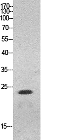
• Western Blot — sharp nuclear band at ~25 kDa; if you see a ladder or smear into higher masses, think HMGB1–protein crosslinks or post-translational adducts (acetylation/SUMO context)
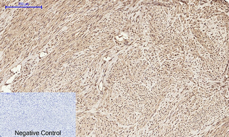
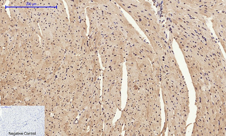
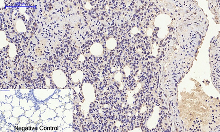
• IHC-P (FFPE) — the defining nuclear staining pattern; loss-of-nuclear → cytoplasmic shift is a visual readout of cellular stress you can map across an entire tissue section
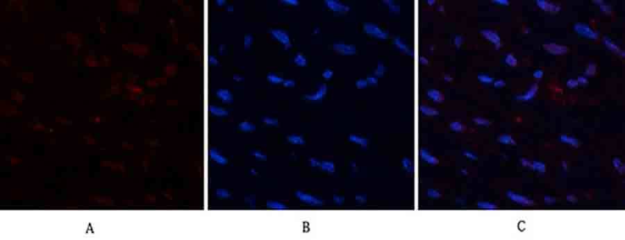
• IF/ICC — co-label with DAPI/RAGE/TLR4/autophagy markers to prove subcellular trafficking
• ELISA (capture/detection) — for quantifying extracellular HMGB1 in supernatant/plasma/serum when paired with a proper redox/complex-disruption prep
Specs you'll typically cite:
• Host / Isotype: Rabbit IgG (polyclonal, affinity purified)
• MW (calculated): ~24.9 kDa (runs ~25 kDa)
• Concentration: ~1 mg/mL, supplied in PBS, pH 7.4, 50% glycerol, 0.5% BSA, 0.09% sodium azide
• Reactivity: Human, Mouse, Rat
• Applications: WB (1:500–2,000 range), IHC-P (1:100–300), IF/ICC (1:50–200), ELISA (suitable as capture or detection in sandwich formats)
The Critical HMGB1 Antibody Pitfall: Nuclear vs. "Extracellular" — They're Not the Same Molecule
A huge fraction of confusing HMGB1 data comes from treating it as if it's a simple on/off secretory cytokine. It's not. The intracellular pool is nuclear, the extracellular pool is often complexed with DNA, histones, LPS, nucleosomes, or CXCL12, and the redox state dictates receptor preference. A few rules keep ABP53233 honest:
- For WB on cells/tissues: run a histone H3 or lamin A/C control alongside — if HMGB1 signal is huge but histones leak, your "HMGB1 translocation" might just be a lysis tear.
- For IHC-P: HMGB1 should be a crisp nuclear signal in healthy zones and show cytoplasmic loss / dropout in necrotic/infarcted cores. Use pH 9.0 EDTA retrieval for FFPE; the nuclear pattern is your internal quality control.
- For extracellular HMGB1 (supernatant/plasma): you're detecting a trace DAMP in a protein-rich soup — either use a sandwich ELISA format or TCA/precipitate + concentrate before blotting. Free extracellular HMGB1 can be pg–low ng/mL; a direct lysate-style WB often won't cut it.
Where This Antibody Earns Its Keep
- Sepsis, trauma, and sterile inflammation (DAMPs)
Quantify/map HMGB1 release in LPS/shock models, hemorrhagic trauma, ischemia–reperfusion (I/R) organ injury (liver, kidney, heart, gut). The canonical story: HMGB1 is a late mediator (peaking 8–24 h) that sustains cytokine storms even after LPS itself clears.
- Autoimmunity & rheumatic disease
HMGB1–autoantibody–nucleosome complexes and RAGE-driven inflammation sit at the center of SLE, RA, dermatomyositis, vasculitis pathogenesis. IHC on synovial/dermal/renal sections with ABP53233 shows you where the alarmin is spilling.
- Cancer & the tumor microenvironment
Tumor cells both overproduce HMGB1 (genomic stress) and secrete it to promote angiogenesis, metastasis, macrophage recruitment, and chemoresistance via RAGE/TLR4. Nuclear-to-cytoplasmic shift in tumor biopsies is a well-recognized histologic correlate of more aggressive behavior.
- Wound healing & tissue repair
Keratinocytes, fibroblasts, and endothelial cells all respond to extracellular HMGB1 (RAGE → MAPK/NF-κB → migration, proliferation, sprouting). IF co-labels (HMGB1 + K14/K10, vWF, α-SMA) let you build the spatial story.
- Neuroinflammation & neurodegeneration
HMGB1 is released by activated microglia and damaged neurons; RAGE binding can drive neuronal stress signaling. Nuclear HMGB1 in NeuN+ cells vs. extracellular HMGB1 in Iba1+ zones is the spatial question ABP53233 was built to answer.
The Bottom Line
HMGB1/HMG-1 is the molecule that taught immunology the word "alarmin" — a chromatin protein that doubles as a danger signal, a DNA chaperone that moonlights as a cytokine, and a nuclear resident that predicts outcome the moment it walks out the door. The HMG-1 Polyclonal Antibody (ABP53233) from Abbkine gives you the multi-platform, rabbit IgG, cross-species (Hu/Mo/Rt) toolset to detect it where it belongs (nucleus) and — just as importantly — document when it doesn't: WB ~25 kDa, crisp IHC-P nuclear pattern, IF for translocation, ELISA-ready for extracellular quantification. In DAMP biology, your antibody isn't just a reagent. It's your witness.
Product Reference: ABP53233 – HMG-1 Polyclonal Antibody
Learn more and order: https://www.abbkine.com/product/hmg-1-polyclonal-antibody-abp53233/


