


The Lactate Gatekeeper: Why Quantifying MCT4 (SLC16A3) Is Essential for Cancer Metabolism, Exercise Physiology, and Beyond — And How the KTE60625 Sandwich ELISA Makes It Routine
Every cell that cranks up glycolysis under hypoxia or proliferative demand faces an existential problem: lactate buildup. If lactic acid accumulates unchecked, intracellular pH crashes, glycolysis stalls, and the cell suffocates in its own waste. The solution is a dedicated proton‑coupled exporter: Monocarboxylate Transporter 4 (MCT4, encoded by SLC16A3). Unlike its relative MCT1, which tends to import lactate for oxidative fuel, MCT4 is the high‑capacity, low‑affinity efflux pump that flushes lactate and H⁺ out of glycolytic cells—making it indispensable in fast‑twitch muscle fibers, activated immune cells, and perhaps most consequentially, cancer cells addicted to aerobic glycolysis (the Warburg effect). In the tumor microenvironment, MCT4 expression on cancer‑associated fibroblasts and hypoxic tumor cells drives the "lactate shuttle" that fuels oxidative tumor regions and acidifies the stroma, promoting invasion and immune escape. Clinically, MCT4 overexpression correlates with poor prognosis in triple‑negative breast cancer, clear‑cell renal cell carcinoma, colorectal cancer, and glioblastoma. Yet despite its centrality, most labs still track MCT4 by mRNA or semi‑quantitative Western blots—tools ill‑suited for the precise, reproducible protein quantification that biomarker validation and drug‑response monitoring demand. The Human Monocarboxylate Transporter 4 (SLC16A3) ELISA Kit (KTE60625) from Abbkine closes that gap, providing a sandwich ELISA platform that turns MCT4 from a "band‑intensity guess" into a calibrated, plate‑readable variable.
MCT4 Biology in Brief: The Lactate Efflux Machine That Shapes Tumors and Muscles
MCT4 is a 12‑transmembrane‑domain protein (~50–55 kDa) belonging to the SLC16 family of proton‑coupled monocarboxylate transporters. Its kinetic signature—Km for L‑lactate ~17–28 mM (much higher than MCT1's ~3–5 mM)—means it only starts working when intracellular lactate piles up, making it the perfect "overflow valve" for highly glycolytic cells. Expression is driven primarily by HIF‑1α under hypoxia, and by MYC and PGC‑1α in certain metabolic contexts.
In cancer, the MCT4 story is especially compelling:
• Hypoxic tumor cells upregulate MCT4 to export lactate, which is then taken up by neighboring oxidative tumor cells via MCT1 and oxidized for ATP—a metabolic symbiosis known as the "lactate shuttle."
• The exported lactate acidifies the extracellular milieu, promoting matrix metalloproteinase activity, angiogenesis, and suppression of antitumor immune cells.
• MCT4 is also highly expressed on cancer‑associated fibroblasts (CAFs), which undergo aerobic glycolysis and feed lactate to epithelial cancer cells.
• Pharmacological inhibition or genetic silencing of MCT4 reduces tumor growth, invasion, and metastasis in preclinical models, establishing it as a validated therapeutic target.
Outside oncology, MCT4 is critical for:
• Skeletal muscle physiology: Fast‑twitch (Type II) fibers rely on MCT4 for lactate clearance during strenuous exercise.
• Placental and retinal metabolism: High‑glycolytic tissues that require rapid lactate export.
• Macrophage polarization: Pro‑inflammatory (M1) macrophages upregulate MCT4 to sustain glycolysis.
Given this breadth, the ability to measure MCT4 protein quantitatively in tissue lysates, cell lysates, serum/plasma (if shed or exosomal), and culture supernatants unlocks a dimension of analysis that mRNA or Westerns cannot reliably provide.
Why a Sandwich ELISA for MCT4? Three Irreplaceable Advantages
- Absolute quantification, not relative density. A sandwich ELISA uses a recombinant MCT4 standard curve on every plate to convert OD₄₅₀ into ng/mL (or pg/mL). This means you can compare MCT4 levels across different experiments, labs, and time points with statistical confidence—something densitometry cannot guarantee.
- Two‑epitope specificity. The pre‑coated capture antibody and the biotinylated detection antibody recognize two distinct epitopes on the MCT4 protein. This double selection dramatically reduces the chance of cross‑reactivity with other monocarboxylate transporters (MCT1, MCT2, MCT3) or unrelated membrane proteins, which is a common pitfall in Western blotting where a single antibody may hit multiple bands.
- High throughput and scalability. A 96‑well plate lets you process up to 40 unknowns (in duplicate) plus a full standard curve in a single run—perfect for dose‑response experiments, time courses, patient cohort comparisons, or screening applications.
Assay Principle: The KTE60625 Sandwich ELISA in a Nutshell
The KTE60625 kit follows the classic, field‑proven two‑site sandwich ELISA architecture:
- A microplate is pre‑coated with a capture antibody specific for human SLC16A3/MCT4.
- Standards and samples (serum, plasma, tissue homogenates, cell lysates, cell culture supernatants, or other biological fluids) are added; MCT4 present in the sample binds to the immobilized antibody.
- After thorough washing, a biotin‑conjugated detection antibody (targeting a different epitope) is added, forming the sandwich complex.
- Streptavidin‑Horseradish Peroxidase (SA‑HRP) binds to the biotin.
- TMB substrate is added; HRP catalyzes the conversion to a blue product, proportional to the amount of captured MCT4.
- Stop solution (acid) turns the solution yellow; absorbance is read at 450 nm.
- Unknown concentrations are interpolated from the MCT4 standard curve run on the same plate.
Total assay time is typically 3–5 hours, depending on incubation steps and user experience.
Performance Specifications That Matter for Publication
Based on the validated parameters for Abbkine's targeted ELISA kits (and consistent with the KTE60625 product family), you can expect:
Parameter Typical Value
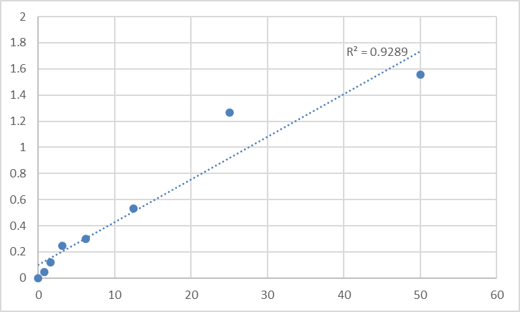
Detection Range 0.156 – 10 ng/mL (7‑point standard curve)
Sensitivity (LOD) ~0.04–0.08 ng/mL
Intra‑Assay CV < 6%
Inter‑Assay CV < 9%
Recovery (spike‑and‑recovery) 92–108% in serum, plasma, and cell lysate matrices
Specificity No significant cross‑reactivity with MCT1, MCT2, MCT3, or other SLC16 family members
Sample Types Validated Serum, plasma (EDTA/heparin), tissue homogenates, cell lysates, cell culture supernatants
Always refer to the lot‑specific certificate of analysis for exact values.
Where Quantifying MCT4 Protein Transforms Your Research
- Cancer Metabolism & Tumor Microenvironment Studies
• Measure MCT4 in tumor lysates or patient biopsies to correlate with hypoxia markers (CA9, HIF‑1α), glycolytic flux (LDHA, PKM2), and clinical outcomes.
• Track MCT4 dynamics in 3D spheroids or organoids under normoxia vs. hypoxia, or after treatment with MCT4 inhibitors (e.g., syrosingopine, AZD3965‑related compounds).
• Quantify MCT4 in CAFs vs. cancer cells from co‑culture systems to dissect the lactate shuttle.
- Exercise & Muscle Physiology
• Monitor MCT4 upregulation in skeletal muscle after endurance training, sprint intervals, or electrical stimulation protocols.
• Correlate MCT4 protein levels with blood lactate clearance rates in animal models or human biopsy specimens.
- Macrophage & Immune Cell Metabolism
• Quantify MCT4 in LPS‑activated macrophages vs. IL‑4‑polarized macrophages to link glycolysis to inflammatory cytokine production.
• Study MCT4 in tumor‑infiltrating lymphocytes (TILs) — lactate export may affect T‑cell persistence and function.
- Drug Development & Target Engagement
• Screen small‑molecule MCT4 inhibitors by measuring the reduction of MCT4 protein in treated cells (or by using the ELISA as a pharmacodynamic readout in xenograft lysates).
• Validate CRISPR‑mediated SLC16A3 knockout or knockdown at the protein level with a calibrated assay instead of a single Western blot.
- Biomarker Discovery
• Explore circulating MCT4 (exosomal or soluble) in plasma from cancer patients as a potential non‑invasive biomarker for tumor glycolytic burden or treatment response.
Practical Tips for Getting Clean MCT4 ELISA Data
• Membrane protein extraction: MCT4 is an integral membrane protein. For cell/tissue lysates, use a RIPA‑style buffer with 1% Triton X‑100 or NP‑40 plus protease inhibitors. Clarify by centrifugation at 12,000–16,000 × g for 15 min at 4°C.
• Normalize to total protein: Always measure total protein (BCA or Bradford) on the same lysate and express MCT4 as ng per mg total protein to control for loading differences.
• Avoid repeated freeze‑thaw: Aliquot samples and standards; MCT4 can degrade with multiple cycles.
• Matrix matching: If you are using serum or plasma, run a blank (sample diluent only) and a known spike to verify recovery in your specific matrix.
• Include a positive control lysate (e.g., HeLa or MDA‑MB‑231 cells cultured under hypoxia) on every plate to monitor inter‑assay consistency.
The Bottom Line
MCT4 is no longer just a footnote in the lactate transporter family—it is a central node in cancer metabolism, muscle physiology, and immune regulation. But to leverage it as a biomarker, a drug target, or a mechanistic readout, you need to measure it with the same rigor you apply to every other quantitative variable in your experiment. The Human Monocarboxylate Transporter 4 (SLC16A3) ELISA Kit (KTE60625) from Abbkine delivers that rigor: a pre‑validated sandwich ELISA with broad dynamic range, excellent specificity, and a workflow that fits into a single afternoon. Whether you are mapping the metabolic landscape of a tumor, training a cohort of mice, or screening the next generation of lactate‑transport inhibitors, this kit turns MCT4 from a fuzzy band into a number you can trust.
Product Reference: KTE60625 – Human Monocarboxylate Transporter 4 (SLC16A3) ELISA Kit
Learn more and order: https://www.abbkine.com/product/human-monocarboxylate-transporter-4-slc16a3-elisa-kit-kte60625/


