


The Inflammation Catabolic That Flies Under Your Radar: Why PTGR1/NADPH-Dependent Prostaglandin Reductase Quantification Completes Your PG/LTB4 Resolution Story
Everyone who works in inflammation knows the headline acts — COX-2, mPGES-1, PGE₂, and maybe PGD₂/J-series if you're deep in resolution biology — but the molecule that actually pulls the plug on prostaglandin signaling at the metabolic endpoint is a compact, membrane-associated oxidoreductase that most people forget to measure: PTGR1, also cataloged as NADPH-dependent prostaglandin reductase 1, LTB4 12-hydroxydehydrogenase, or prostaglandin 15-dehydrogenase (NADP+)–dependent (gene PTGR1, UniProt: P15428, ~36–38 kDa, 328 aa). PTGR1 lives predominantly in the endoplasmic reticulum and nuclear envelope, where it uses NADPH to reduce/oxidize prostaglandin and eicosanoid carbonyl groups (including PGE₂ → 15-keto-PGE₂ / PGD₂ catabolism) and contributes to the oxidative inactivation of LTB4 (→ 12-oxo-LTB4 → further catabolism) — in short, it is one of the enzymes that turns active eicosanoids into their inactive/less-active keto-forms, helping the cell exit the inflammatory loop. The Human Prostaglandin reductase 1 (PTGR1) ELISA Kit (KTE61068) from Abbkine is designed to finally give you a quantitative plate-readout of the enzyme protein itself, so your prostaglandin / eicosanoid resolution narrative isn't just a panel of LC-MS peaks and ELISA for PGE₂ — it's also the catabolic capacity that determines whether those mediators accumulate or get swept away.
PTGR1 in Context: The Metabolic "Off-Switch" for Bioactive Lipids
The cyclooxygenase pathway pumps out PGG₂ → PGH₂ → a suite of bioactive prostanoids (PGE₂, PGD₂, PGI₂, TXA₂) that drive pain, fever, vasodilation/constriction, platelet aggregation, and stromal inflammation. But every active mediator also needs a defined clearance and deactivation track, and that's where PTGR1 enters:
• PTGR1 (NADPH-dependent) performs carbonyl reduction/oxidation reactions on the prostaglandin cyclopentenone core that attenuate biological potency and mark prostanoids for further catabolism and excretion.
• It overlaps functionally with the better-known cytosolic 15-PGDH (HPGD/NAD+-dependent) but is distinguished by its NADP(H) preference, ER/nuclear envelope locale, and its broader activity on substrates such as the J-series (PGJ₂, 15d-PGJ₂) and LTB4-related catabolism.
• Expression is regulated by carcinogen exposure, oxidative stress, hypoxia, and steroid signaling, and PTGR1 has been implicated both as a tumor suppressor–associated catabolite (loss → prostanoid excess → tumor-promoting inflammation) and as a pro-resolving adaptation (upregulation → helps terminate eicosanoid signaling).
Put simply: if you're measuring how much PGE₂ or LTB4 is being made, the scientifically honest next question is "how fast can this cell turn it off?" — and PTGR1 protein level is a core part of that answer.
Why a Sandwich ELISA for PTGR1 — And Why Gel-Based "Presence" Isn't Enough
PTGR1 is ~37 kDa, non-secreted, and ER/nuclear envelope–associated, which means:
- You need good membrane/particulate prep (or at least clarified whole-cell lysate) to see a clean signal — crude cytosolic-only spins can undercount it.
- It's not a high-abundance housekeeper, so fold-changes matter and a densitometric band with 3 lanes is rarely defensible as a quantification.
- Specificity matters: the reductase family has related members (HPGD/15-PGDH, PTGR2/CBR1 crossing over in some assays), so a two-epitope sandwich ELISA is the rigorous way to lock it down.
The KTE61068 kit delivers exactly that architecture:
- Pre-coated microplate with a capture antibody specific for human PTGR1.
- Standards + samples (tissue homogenates, cell lysates, microsomal fractions, other biological fluids) added → PTGR1 binds.
- Wash → biotinylated anti-PTGR1 detection antibody (different epitope) forms sandwich.
- Streptavidin–HRP → TMB → color ∝ bound PTGR1.
- Stop → read 450 nm → interpolate PTGR1 concentration from the standard curve.
Performance Snapshot (the numbers you'll cite)
Parameter Typical KTE61068-class spec
Target Human PTGR1 (UniProt P15428, PTGR1 / LTB4DH)
Format 96-well sandwich ELISA, pre-coated capture
Detection Biotin-Ab → SA-HRP → TMB, 450 nm
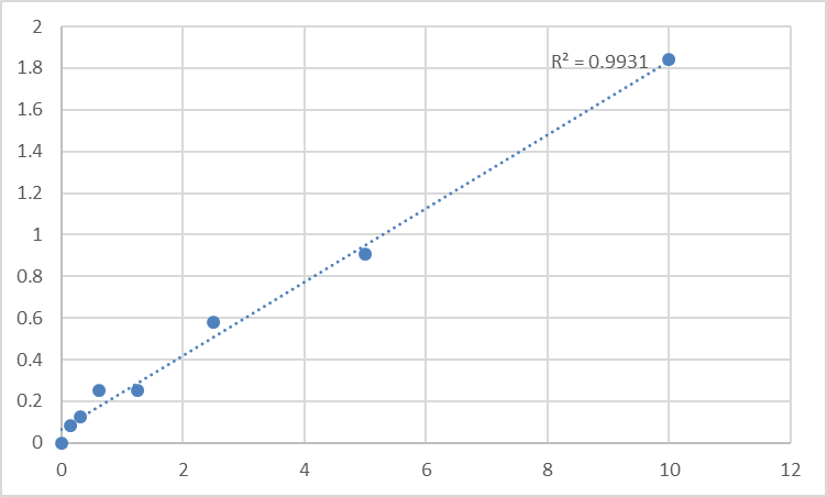
Dynamic Range 0.156 – 10 ng/mL (7-point standard)
Sensitivity / LOD ~0.07–0.10 ng/mL
Intra-Assay CV < 8%
Inter-Assay CV < 10–12%
Samples Tissue homogenates, cell lysates, cell culture supernatants/lysates, other biological fluids
Assay time ~3–5 hours
(Always confirm exact range, dilution scheme, and lot-specific recovery on the shipped CoA/datasheet.)
Where Quantifying PTGR1 Protein Actually Completes the Eicosanoid Story
- NSAID / COX-2 / mPGES-1 drug discovery & toxicology
You can show "PGE₂ dropped" with an LC-MS or PGE₂ ELISA — but adding PTGR1 protein (ng/mg total protein) lets you distinguish two very different mechanisms:
• Drug → less PGE₂ made (upstream COX/mPGES block)
• Drug → PGE₂ still made, but catabolism up (PTGR1/HPGD induction = pro-resolving shift)
That distinction is why journals like the resolution field now expect at least a nod to catabolic enzymes alongside the prostanoid readouts.
- Cancer inflammation & tumor microenvironment
Many tumors suppress PTGR1/HPGD catabolism (via hypermethylation, oxidative stress adaptation, or miRNA regulation), allowing a pro-tumor PGE₂/EP signaling loop to persist. Measuring PTGR1 in tumor lysates vs. adjacent tissue (normalized to total protein or epithelial/stromal markers) gives you the catabolic-capacity axis that PGE₂ ELISA alone can't resolve.
- Lung inflammation, asthma & LTB4 axis
Because PTGR1 contributes to LTB4 oxidative inactivation (12-OH → 12-oxo), its expression in alveolar macrophages, bronchial epithelium, and lung parenchyma matters for whether LTB4 signals resolve or linger. A plate-readout scales across BAL-cell lysates or lung-tissue homogenates far better than a gel-only approach.
- Oxidative stress & xenobiotic models
PTGR1 also has quinone reductase-like activity (detox of certain α,β-unsaturated carbonyls), so it can move with NRF2/ARE induction — quantifying it ties your ARE-reporter or NRF2-KD experiment to a real enzymatic protein, not just a DCF fluo信号.
- CRISPR/siRNA & rescue validation
Knocking PTGR1 out? Don't just show "band fainter." Report % PTGR1 protein remaining ± SEM from a calibrated curve, and — if your story allows — pair with PGE₂ / PGD₂ / 15d-PGJ₂ metabolite profiling so the enzyme loss has a functional lipid consequence.
Quick Prep Note (PTGR1 Is Particulate — Treat It That Way)
• Lyse in cold buffer with 0.5–1% Triton X-100 or NP-40 + protease inhibitors; a post-nuclear spin (10,000–16,000 ×g, 4°C, 15 min) clears debris, and the supernatant contains ER/nuclear envelope membrane fragments where PTGR1 lives.
• BCA the same lysate → report ng PTGR1 / mg total protein.
• Aliquot, avoid >2 freeze–thaw cycles, warm all kit reagents to RT ≥ 30 min before opening, protect TMB from light, stop uniformly, read 450 nm promptly, and run the full standard curve on every plate.
The Bottom Line
PTGR1 is the ER-localized catabolic workhorse that helps terminate prostaglandin and leukotriene signaling — the metabolic "off-switch" that makes your "PGE₂ is high" claim meaningful by defining the cell's capacity to clear it. The Human Prostaglandin reductase 1 (PTGR1) ELISA Kit — KTE61068 from Abbkine gives you a pre-coated sandwich ELISA to quantify that off-switch as an interpolated concentration, not a guess: capture → biotin detection → HRP–TMB → 450 nm → ng/mL, in a ~3–5 hour workflow that fits inside a real lab day and produces numbers you can normalize, replicate, and defend.
Product Reference: KTE61068 – Human Prostaglandin reductase 1 (PTGR1) ELISA Kit
Learn more and order: https://www.abbkine.com/product/human-prostaglandin-reductase-1-ptgr1-elisa-kit-kte61068/
(For Research Use Only; not for diagnostic procedures in humans.)


