


The Collagen Breaker That Opens the Tumor Highway: Why Quantifying MMP-1 Protein — Not Just Its Activity — Changes How You Read Invasion, Fibrosis, and Plaque Rupture
Every tissue in your body depends on collagen for structural integrity — and that means every tissue also needs a precise, regulated way to cut it. Enter Matrix Metalloproteinase-1 (MMP1), better known as interstitial collagenase or collagenase-1: the founding member of the MMP family and the only enzyme in human biology capable of cleaving the triple-helical domain of native types I, II, and III fibrillar collagen at physiological pH. It performs that legendary single-stranded nick three-quarters of the way down the helix, turning an indestructible cable into denatured gelatine-prone fragments — and once that barrier falls, the rest of the degradative machinery (MMP-2, MMP-9, MT1-MMP) moves in to finish the job. This is why MMP-1 sits at the very center of embryonic morphogenesis, uterine remodeling, bone resorption, wound healing, and aneurysm-prone vascular disease — and why, when its control fails, it becomes the proverbial skeleton key for cancer cell invasion, atherosclerotic plaque cap thinning, rheumatoid pannus erosion, and pulmonary emphysema destruction. The problem is that most labs still treat MMP-1 as a "zymography band" or an indirect activity assay, when what the experiment actually demands is a specific, protein-level, plate-readable concentration. The Human Matrix Metalloproteinase 1 (MMP1) ELISA Kit (KTE61122) from Abbkine is built to give you exactly that: a quantitative sandwich ELISA that reads total MMP-1 (pro-form + active) so your invasion, remodeling, or fibrosis story rests on a number — not a gelatin gel shadow.
MMP-1 101: The Interstitial Collagenase That Defines "Remodeling" vs. "Destruction"
MMP1 (UniProt: P03956, gene MMP1, 469 aa precursor) is synthesized as a pre-pro-enzyme (54–57 kDa) that loses its N-terminal signal peptide and becomes the pro-MMP1 zymogen (51–53 kDa), kept quiescent by its cysteine-switch motif (PRCGXPD…) where a conserved Cys coordinates the catalytic Zn²⁺ to block substrate access. Activation — primarily by plasmin (generated from plasminogen via uPA/tPA at the cell surface) or by MT1-MMP (MMP14) — cleaves the pro-domain, exposing the active site and yielding the mature active MMP-1 (42–45 kDa, glycosylation-dependent). Its natural brake is the tight-binding inhibitor TIMP-1 (formed in near-stoichiometric opposition), so the biologically relevant readout in any system is never just "[MMP-1]" in isolation — it's the balance of active MMP-1 vs. TIMP-1, and whether pro-MMP-1 is being converted at the right place and time.
The substrates that matter most:
• Collagen I, II, III — the triple helices themselves (unique to interstitial collagenases)
• Entactin/nidogen, aggrecan, periostin, and multiple non-collagen ECM proteins as secondary targets
• Pro-MMP-9 and pro-MMP-13 — cross-activation (MMP-1 can help prime other MMPs)
Why a Sandwich ELISA for MMP-1 — And Why Zymography Alone Won't Carry Your Claim
Gelatin zymography is beautiful for showing enzyme activity as a band, but it has three structural weaknesses for modern quantitative claims:
- It measures catalytic function, not protein mass — an active form and a partially inhibited form can differ at the protein level while looking "similar" on a gel, and pro-enzyme often doesn't enter the activity lane at all.
- It's semi-quantitative at best — densitometry across a 10-lane gel with variable background is not a substitute for a calibrated standard curve.
- It can't easily discriminate MMP-1 from MMP-8/MMP-13 in complex samples without very specific inhibition or antibody overlay — and even then you're inferring identity.
A sandwich ELISA bypasses these by using two independent anti-MMP-1 epitopes (pre-coated capture + biotin detection) → Streptavidin–HRP → TMB → 450 nm → interpolated ng/mL from a recombinant MMP-1 standard curve. You get total MMP-1 protein (pro + active pool, unless you specifically immunodeplete/separate), and you get it in a format that scales to time courses, dose-responses, and clinical-cohort plates without chaining yourself to a zymogram washer.
Assay Principle: KTE61122 Sandwich ELISA
- A microplate is pre-coated with a capture antibody specific for human MMP-1.
- Standards and samples (serum, plasma, tissue homogenates, cell lysates, cell culture supernatants, other biological fluids) are added; MMP-1 present binds.
- After washing, a biotinylated anti-MMP-1 detection antibody (different epitope) forms the sandwich complex.
- Streptavidin–HRP binds biotin → TMB → color ∝ bound MMP-1.
- Stop solution → yellow → read Absorbance at 450 nm → interpolate unknowns from the MMP-1 standard curve.
Typical performance envelope for this kit family (always confirm lot-specific values on the Abbkine datasheet):
Parameter Typical Specification
Target Human MMP-1 (collagenase-1, UniProt P03956)
Format 96-well sandwich ELISA, pre-coated capture
Detection Biotin-Ab → SA-HRP → TMB, 450 nm read
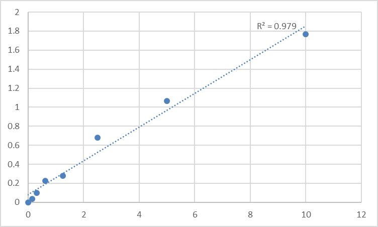
Dynamic Range 0.156 – 10 ng/mL
Sensitivity / LOD ~0.045–0.08 ng/mL
Intra-Assay CV < 7–8%
Inter-Assay CV < 10%
Specificity No significant cross-reactivity with other MMPs (MMP-8, MMP-13) at physiological levels
Samples Serum, plasma (citrate/EDTA preferred for MMP work), tissue homogenates, cell lysates, culture supernatants
Assay time ~3–5 hours
Where Quantifying MMP-1 Protein Actually Moves the Needle
- Cancer Invasion & Metastasis (the "stromal breach" axis)
MMP-1 is the enzyme that cuts the collagen I wall around a tumor nest, enabling collective invasion and single-cell dissemination. In fibroblasts and cancer cells, its expression is driven by TNF-α, IL-1β, AP-1 (c-Fos/c-Jun), and ETS transcription factors, and it is restrained by TIMP-1, TGF-β (paradoxical in some contexts), and steroid hormones. ELISA-quantified MMP-1 in conditioned media, tumor lysates, or (exploratorily) plasma gives you the protein mass that links transcriptional hits to ECM destruction — far more defensible than an n=3 zymogram.
- Arthritis & Osteoarthritis (cartilage and synovial erosion)
Synovial fibroblasts and chondrocytes under pro-inflammatory cues secrete MMP-1 to degrade type II/I collagen in the articular matrix; measuring it in synovial fluid, synovial tissue lysates, or culture supernatants from RA/OA models is the direct readout of erosive protease burden (again, best paired with TIMP-1 to get the ratio).
- Atherosclerosis & Plaque Vulnerability
The fibrous cap of an advanced coronary plaque is a collagen-I-dense scar — and its thinning is an MMP story. Elevated MMP-1 (and MMP-9, MT1-MMP) vs. TIMP-1 shifts the cap toward rupture risk. Being able to quantify MMP-1 from plaques, carotid endarterectomy tissue lysates, or even exploratory plasma adds a structural-dynamics variable to your lipid/inflammation panel.
- Pulmonary Emphysema & COPD (collagen I/III destruction in lung parenchyma)
Smoking and chronic protease–antiprotease imbalance (elastase/MMP vs. α₁-AT) drive alveolar wall loss — MMP-1 is a direct effector of the collagen framework destruction. Quantifying it in BAL fluid, lung tissue homogenates, or macrophage lysates ties the smoking/oxidative-stress signal to a specific collagenase, not just "total protease activity."
- Wound Healing & Fibrosis (too much vs. too little)
Both extremes are bad: insufficient MMP-1 → chronic non-healing ulcers; excessive/unresolved MMP-1 → hypertrophic scar, destructive remodeling, or fibrotic progression. The ELISA lets you track MMP-1:TIMP-1 dynamics across granulation tissue timepoints instead of guessing.
- CRISPR/siRNA & drug screens (MMP modulators)
Testing MMP-1 siRNA, TIMP-1 overexpression, uPA/plasmin axis inhibitors, or MAPK/AP-1 pathway drugs? Report % MMP-1 protein remaining ± SEM from a calibrated curve, normalized to mg total protein (BCA). Reviewers notice the difference between "band lighter" and "4.6 ng/mg reduced, p < 0.01."
A Quick Prep/Handling Note (Because MMPs Are Finicky in Blood)
• Citrate or EDTA plasma is preferred over serum for many MMP studies — clot formation itself can activate platelets/uPA/plasmin and artifactually spike MMP levels. Process cold, spin promptly, aliquot, store -80°C, avoid repeated freeze–thaw.
• For tissue/cell lysates: lyse in cold RIPA or Tris–Triton (0.5–1% NP-40) + protease inhibitors + 5–10 mM Ca²⁺ (MMP catalytic domain likes the right metal), clarify by centrifugation, keep on ice, run BCA on the same final lysate → express as ng MMP-1 / mg total protein.
• Run the full standard curve on every plate — MMP-1 adsorption quirks mean plate-to-plate drift is real, and the curve is your insurance.
The Bottom Line
MMP-1 is the enzyme that decides whether fibrillar collagen is a wall or a doorway — and when that decision goes wrong, it writes the pathology of invasion, erosion, and rupture across cancer, arthritis, vascular disease, and lung destruction. Measuring it deserves more than a gelatin gel and a densitometry guess. The Human Matrix Metalloproteinase 1 (MMP1) ELISA Kit — KTE61122 from Abbkine gives you the right tool for the job: pre-coated capture → biotin detection → HRP–TMB → 450 nm → ng/mL, in a ~3–5 hour workflow that scales from a 6-well time-course to a 50-sample cohort without sacrificing specificity. When your paper's hinge-point is "collagen was degraded," make sure your evidence hinge-point is a number, not a shadow.
Product Reference: KTE61122 – Human Matrix Metalloproteinase 1 (MMP1) ELISA Kit
Learn more and order: https://www.abbkine.com/product/human-matrix-metalloproteinase-1-mmp1-elisa-kit-kte61122/
(For Research Use Only; not for diagnostic procedures in humans.)


