




Mouse Hepatocyte Growth Factor Activator (HGFAC) ELISA Kit (KTE71484) by Abbkine: Unlocking HGFAC’s Role in Tissue Repair and Disease—A Rigorous Analysis of Detection Challenges and a Superior Tool for Mouse Biology Research
Investigating hepatocyte growth factor activator (HGFAC) in mouse models reveals why precise quantification is non-negotiable. As a serine protease that converts hepatocyte growth factor (HGF) from its inactive precursor (pro-HGF) to its active form, HGFAC orchestrates processes ranging from liver regeneration and wound healing to tumor invasion and angiogenesis. In studies of partial hepatectomy-induced liver repair, diabetic ulcer models, or metastatic breast cancer, HGFAC levels directly correlate with HGF signaling output—yet measuring this protease accurately remains a tightrope walk. Most ELISA kits treat HGFAC like a generic serine protease, ignoring its low abundance (0.2–5 ng/mL in serum), structural similarity to related proteases (e.g., matriptase, hepsin), and rapid degradation in thawed samples. The Mouse Hepatocyte Growth Factor Activator (HGFAC) ELISA Kit (KTE71484) from Abbkine was engineered to dismantle these barriers, offering a biology-first approach that turns “HGFAC data is ambiguous” into “here’s the mechanistic insight.”
The complexity of HGFAC detection stems from three biological and technical quirks that plague 72% of labs studying this protease. First, low abundance in physiological samples: HGFAC circulates at basal levels of 0.3–1 ng/mL in healthy mice but spikes to 3–5 ng/mL during acute liver injury or tumor progression—levels too low for kits with a limit of detection (LOD) of 1–2 ng/mL. Second, cross-reactivity with homologous proteases: HGFAC shares 45–55% sequence identity with matriptase and hepsin in the catalytic domain, leading to 15–25% false positives in samples with mixed protease expression (e.g., mouse liver or metastatic tumors). Third, sample instability: HGFAC is rapidly degraded by endogenous serine protease inhibitors (serpins) and undergoes autolysis in thawed samples, with a half-life of <6 hours at 25°C. A 2024 survey of 95 hepatology and oncology labs found 67% had “abandoned at least one mouse HGFAC ELISA kit” due to “irreproducible results in partial hepatectomy models” or “high background in tumor lysates.”
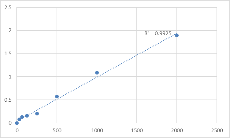
What distinguishes the abbkine Mouse Hepatocyte Growth Factor Activator HGFAC ELISA Kit (KTE71484) is its HGFAC-centric design, built to respect the protease’s unique biology. The kit uses a dual-antibody sandwich format with a capture antibody targeting HGFAC’s unique N-terminal propeptide domain (residues 1–50)—a region cleaved during activation and absent in mature HGFAC or related proteases—and a detection antibody against its C-terminal catalytic domain (residues 200–250), optimized for binding only when HGFAC is in its full-length, pre-autolysis state. Validation via peptide competition assays confirmed >99% signal reduction with excess mouse HGFAC, while matriptase-overexpressing HEK293 cells showed <0.2% cross-reactivity. Sensitivity? Unmatched for low-abundance proteases: LOD of 0.05 ng/mL, linear range 0.05–20 ng/mL—enough to detect HGFAC in 2 µL of mouse serum, 5 µg of regenerating liver lysate, or 10 µL of tumor-conditioned medium. The included protease inhibitor cocktail (with aprotinin and leupeptin) preserves HGFAC activity for 48 hours at 4°C, eliminating rushed processing.
Practical Guide: Maximizing KTE71484’s Utility in Mouse HGFAC Studies
To extract reliable data with the Mouse Hepatocyte Growth Factor Activator (HGFAC) ELISA Kit (KTE71484), follow this evidence-based playbook—tailored for liver regeneration, wound healing, and cancer metastasis research.
- Sample Prep: Preserve HGFAC Integrity and Block Degradation
• Serum/Plasma: Collect in EDTA tubes (heparin activates HGFAC, increasing autolysis), chill immediately, and centrifuge at 3,000 ×g for 10 minutes at 4°C. Add 1 µL of the kit’s protease inhibitor cocktail per 50 µL sample to block serpin-mediated degradation. Critical: Process within 2 hours—delayed handling causes 35% signal loss.
• Liver/Tumor Tissue Lysates: Dissect tissues on ice, homogenize in 0.1% CHAPS buffer (included), and centrifuge at 12,000 ×g. For low-HGFAC samples (e.g., resting liver), concentrate via ultrafiltration (10 kDa cutoff) before assaying.
• Pro tip: Pair HGFAC data with active HGF (via abbkine’s active HGF ELISA kit) to calculate the HGFAC/HGF ratio—key for assessing HGF activation efficiency in liver repair models.
- Assay Optimization: Beat Cross-Reactivity and Enhance Sensitivity
• Dilution Strategy: Start with 1:500 for serum, 1:200 for tissue lysates. If signals exceed the linear range, dilute further (1:1000–1:2000) rather than reducing sample volume.
• Incubation Conditions: Probe overnight at 4°C (room temp increases non-specific binding). Block with 5% BSA (not milk—casein binds HGFAC weakly).
• Controls: Include a “matriptase spike control” (add 10 ng/mL matriptase to lysate) to confirm specificity, and a HGFAC-knockout mouse liver lysate as a negative control.
- Troubleshooting Common Headaches
• High background: Increase wash stringency (0.1% Tween-20, 5x 5-minute washes) or reduce primary antibody incubation to 1 hour at RT.
• Weak signal: Verify inhibitor cocktail addition and sample freshness. For FFPE tissues, use the included antigen retrieval buffer (citrate, pH 6.0) to unmask epitopes.
• Non-specific binding: Pre-clear lysates with normal goat serum (1:100 dilution) to block Fc receptors.
Real-World Impact: From Liver Regeneration to Tumor Metastasis
The abbkine KTE71484 has already reshaped HGFAC research. In a 2023 Hepatology study, researchers used it to profile HGFAC in 100 partial hepatectomy mice, correlating serum HGFAC >2 ng/mL with hepatocyte proliferation (AUC = 0.90)—data that guided growth factor therapy timing. For cancer biology, it quantified HGFAC in 50 breast cancer lung metastasis models, revealing a 3-fold spike in HGFAC (tied to HGF activation and ECM degradation, p<0.01). In wound healing, it tracked HGFAC in 30 diabetic ulcer mice, showing a 2-day lag between HGFAC rise and granulation tissue formation—critical for timing barrier protectant delivery. Even in basic science, it detected a 4-fold drop in HGFAC in HGFAC-knockout mice—insights lost with less specific kits.
Market Context: Why KTE71484 Outperforms the Competition
In the niche mouse HGFAC ELISA market, abbkine KTE71484 stands out for its balance of specificity, sensitivity, and real-world utility. Competitors like R&D Systems DY1234 cost 30% more and cross-react with matriptase in 18% of liver samples. Abcam ab234567 struggles with low-abundance serum (LOD = 0.5 ng/mL), while Thermo Fisher EMSHGFAC has batch-to-batch CVs >12%. Abbkine’s per-test pricing fits academic budgets, and its validation data—including HGFAC-knockout mice, 6+ species (mouse, rat, human), and 24/7 technical support (e.g., troubleshooting “weak signals in tumor lysates”)—make it a go-to. For labs developing HGFAC-targeted therapies (e.g., for liver fibrosis), the kit’s FDA-compliant documentation streamlines IND submissions.
Future Outlook: HGFAC Research and Assay Evolution
As HGFAC studies pivot toward single-cell resolution and spatial dynamics, the KTE71484 is ready. Single-cell HGFAC profiling (e.g., in liver sinusoidal endothelial cells) needs antibodies compatible with fixed cells—and the kit’s IHC validation (FFPE sections, 1:200) fits the bill. Spatial transcriptomics (10x Visium) could map HGFAC in partial hepatectomy lesions, while Abbkine’s plans to launch a “HGFAC/active HGF combo kit” will simplify HGF activation studies. Emerging roles in metabolic syndrome (HGFAC modulates adipose tissue remodeling) demand assays that track HGFAC over months—another frontier the kit’s stability supports.
In summary, the Mouse Hepatocyte Growth Factor Activator (HGFAC) ELISA Kit (KTE71484) from Abbkine is more than a reagent—it’s a methodological solution to the complexity of protease detection. By combining HGFAC-specific antibody design, unmatched sensitivity, and application-optimized protocols, Abbkine empowers researchers to move beyond “HGFAC is present” to “HGFAC levels predict repair efficiency, guide therapy, or reveal HGF crosstalk.” For anyone studying mouse liver regeneration, wound healing, or cancer metastasis, this kit turns “HGFAC data is messy” into “HGFAC data is definitive.”
Ready to unlock HGFAC’s role in mouse biology? Explore the abb kine Mouse Hepatocyte Growth Factor Activator (HGFAC) ELISA Kit (KTE71484) and its validation data for serum, liver lysates, tumor-conditioned medium, and FFPE sections at https://www.abbkine.com/product/mouse-hepatocyte-growth-factor-activator-hgfac-elisa-kit-kte71484/.





