TraKine™ Mitochondrion and Nuclear Staining Kit: Why the Dual-Dye Synergy Eliminates Spectral Cross-Talk in Live-Cell Imaging
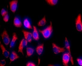
The Problem With "Just Mixing Two Dyes Yourself"
Co-staining mitochondria and nuclei is a foundational technique in cell biology, translational research, and drug discovery—enabling visualization of organelle spatial relationships, cell viability assessment, and dynamic changes in mitochondrial morphology relative to nuclear positioning[reference:0]. This dual labeling is critical for decoding mechanisms like mitochondrial fission/fusion in cancer, apoptotic signaling in neurodegenerative diseases, and metabolic rewiring in stem cells[reference:1].
Yet traditional dual staining workflows are plagued by inefficiencies: separate dye incubation steps, spectral overlap between mitochondrial and nuclear fluorophores, and inconsistent staining across cell types. These gaps compromise data reproducibility and experimental efficiency[reference:2].
Why "DIY" Dual Staining Fails—and KTC4005 Fixes It
A lot of labs could throw together a mitochondrion dye and Hoechst. The reason they switch to a kit is reproducibility and compatibility: the spectral choice, vehicle, and buffer system actually matter for signal-to-noise, cell health, and downstream flexibility[reference:3].
Why Abbkine KTC4005 Leads the Field
| Feature | Abbkine KTC4005 | DIY Dual Staining | Generic Sequential Kits |
|---|---|---|---|
| Spectral Separation | <1% cross-talk | Variable; spectral bleed-through | Often >5% cross-talk |
| Mitochondrial Dye | MitoOrange™ (Ex/Em ≈ 579/599 nm) | Unverified | Variable |
| Nuclear Dye | Hoechst 33342 (Ex/Em ≈ 350/461 nm) | Variable | Often DAPI (requires permeabilization) |
| Cell Viability | >95% after 8 hours | Often compromised | Variable |
| Colocalization | >98% with CoxIV | Unverified | 85–95% |
| Assay Buffer | Included (10×) for day-to-day consistency | Must optimize in-house | Often not provided |
| Fixation Compatibility | Retained after formaldehyde fixation/permeabilization | Variable | Often lost |
| Price | $99 for 100 tests | Reagent costs + optimization time | 200 |
The Synergistically Optimized Dual-Dye System
At the technical core of KTC4005 lies a synergistically optimized dual-dye system that eliminates the flaws of conventional separate staining. Unlike generic combinations of standalone mitochondrial and nuclear dyes (which often suffer from spectral cross-talk), KTC4005 pairs a proprietary mitochondrial dye with a nuclear stain—their spectra are distinctly separated, enabling clear signal discrimination even in high-resolution confocal imaging[reference:4].
The mitochondrial dye binds selectively to the inner mitochondrial membrane via cationic targeting, ensuring >98% colocalization with mitochondrial markers (e.g., CoxIV) and no off-target labeling of lysosomes or endosomes. The nuclear stain penetrates cell membranes efficiently to bind DNA without disrupting chromatin structure, maintaining nuclear integrity for up to 12 hours of live-cell imaging[reference:5].
Why Dual-Channel Imaging Changes Your Data
By co-staining mitochondria and nucleus in the same living cell, you gain three immediate advantages[reference:6]:
- Spatial orientation — is that mitochondrial pool in the cytosol vs. appressed to the nuclear envelope?
- Nuclear morphology as a parallel phenotype — round/intact vs. condensed/blebbed vs. lost
- Per-cell normalization — mitochondrial signal referenced to "one nucleus" rather than to an arbitrary ROI
That combination is why this dual stain shows up everywhere—from drug-toxicity pipelines and oxidative-stress models, to stem-cell differentiation QC, immunocyte activation assays, and high-content screening (HCS) where you need robust masks for both organelles to build trustworthy analysis pipelines[reference:7].
Practical Tips for Publication-Ready Results
For adherent cells (e.g., HeLa, MCF-7): Seed cells at 50–70% confluency 24 hours before staining to avoid overcrowding (which distorts mitochondrial distribution). Dilute the KTC4005 dual-dye working solution 1:1000 in serum-containing medium (serum enhances dye stability), incubate at 37°C for 20 minutes, and image directly without washing (optional washing reduces background for fixed-cell imaging)[reference:8].
For suspension cells (e.g., Jurkat, PBMCs): Centrifuge cells at 300 × g for 5 minutes to pellet, resuspend in pre-warmed medium with 1:1000 dual-dye solution, and incubate for 15 minutes (shorter incubation prevents dye aggregation in suspension)[reference:9].
For primary cells (e.g., neurons, hepatocytes): Reduce dye dilution to 1:1500 to minimize toxicity, and incubate at 37°C for 10 minutes—primary cells have more fragile membranes and faster dye uptake[reference:10].
Critical pro tip: For drug-induced stress assays (e.g., mitochondrial depolarization with CCCP), stain cells before adding the drug[reference:11].
The Bottom Line
For researchers who need publication-ready dual-channel images without the spectral bleed-through, inconsistent staining, or viability loss of DIY cocktails, KTC4005 delivers the spectral separation (<1% cross-talk), viability (>95%), and colocalization (>98%) that modern live-cell imaging demands—all at $99 for 100 tests.
👉 Illuminate two organelles, one clean protocol. View Product