| Product name | c-Myc Polyclonal Antibody |
| Immunogen | Synthesized peptide derived from the C-terminal region of human c-Myc at AA range: 360-440 |
| Host | Rabbit |
| Reactivity | Human,Mouse,Rat |
| Applications | WB,IHC,IF,ELISA |
| Applications notes | Optimal working dilutions should be determined experimentally by the investigator. Suggested starting dilutions are as follows: WB 1:500-1:2000;IHC 1:100-1:300;IF 1:200-1:1000;ELISA 1:40000;Not yet tested in other applications. |
| Clonality | Polyclonal |
| Preparation method | The antibody was affinity-purified from rabbit antiserum by affinity-chromatography using epitope-specific immunogen. |
| Alternative | MYC; BHLHE39; Myc proto-oncogene protein; Class E basic helix-loop-helix protein 39; bHLHe39; Proto-oncogene c-Myc; Transcription factor p64 |
| Formulation | Liquid solution |
| Concentration | 1 mg/ml |
| Molecular weight | 50kD(~60kD in some samples) |
| Storage buffer | Liquid in PBS containing 50% glycerol, 0.5% BSA and 0.02% sodium azide. |
| Storage instructions | Stable for one year at -20°C from date of shipment. For maximum recovery of product, centrifuge the original vial after thawing and prior to removing the cap. Aliquot to avoid repeated freezing and thawing. |
| Shipping | Gel pack with blue ice. |
| Precautions | The product listed herein is for research use only and is not intended for use in human or clinical diagnosis. Suggested applications of our products are not recommendations to use our products in violation of any patent or as a license. We cannot be responsible for patent infringements or other violations that may occur with the use of this product. |
| Background | V-myc avian myelocytomatosis viral oncogene homolog encoded by MYC is a multifunctional, nuclear phosphoprotein that plays a role in cell cycle progression, apoptosis and cellular transformation. It functions as a transcription factor that regulates transcription of specific target genes. Mutations, overexpression, rearrangement and translocation of this gene have been associated with a variety of hematopoietic tumors, leukemias and lymphomas, including Burkitt lymphoma. There is evidence to show that alternative translation initiations from an upstream, in-frame non-AUG (CUG) and a downstream AUG start site result in the production of two isoforms with distinct N-termini. The synthesis of non-AUG initiated protein is suppressed in Burkitt's lymphomas, suggesting its importance in the normal function of MYC. |
| Gene ID | 4609 |
| Alternative | MYC; BHLHE39; Myc proto-oncogene protein; Class E basic helix-loop-helix protein 39; bHLHe39; Proto-oncogene c-Myc; Transcription factor p64 |
| Others | c-Myc Polyclonal Antibody detects endogenous levels of c-Myc protein. |
| Accession | P01106 |
| Observed Band(KD) | 50,(also ~60KD in some samples) |
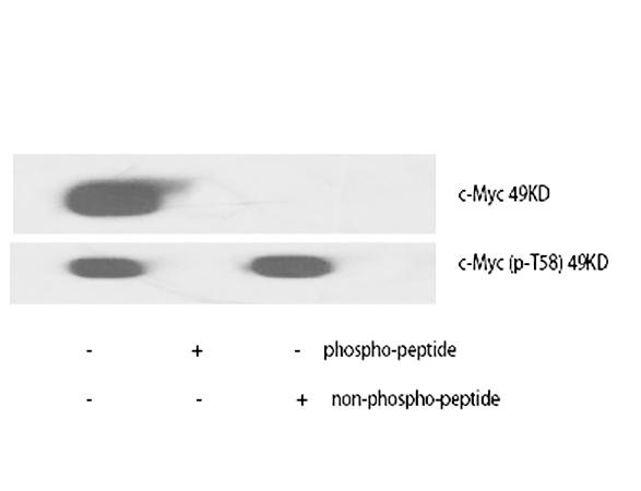
Fig.1. Western Blot analysis of various cells using c-Myc Polyclonal Antibody diluted at 1:1000.
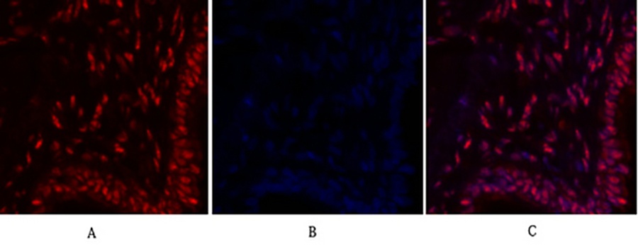
Fig.2. Immunofluorescence analysis of human lung tissue. 1, c-Myc Polyclonal Antibody (red) was diluted at 1:200 (4°C, overnight). 2, Cy3 labeled secondary antibody was diluted at 1:300 (room temperature, 50min). 3, Picture B: DAPI (blue) 10min. Picture A: Target. Picture B: DAPI. Picture C: merge of A+B.
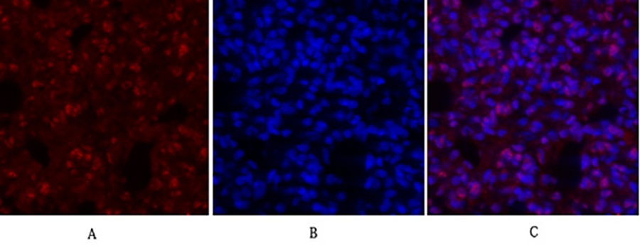
Fig.3. Immunofluorescence analysis of rat lung tissue. 1, c-Myc Polyclonal Antibody (red) was diluted at 1:200 (4°C, overnight). 2, Cy3 labeled secondary antibody was diluted at 1:300 (room temperature, 50min). 3, Picture B: DAPI (blue) 10min. Picture A: Target. Picture B: DAPI. Picture C: merge of A+B.
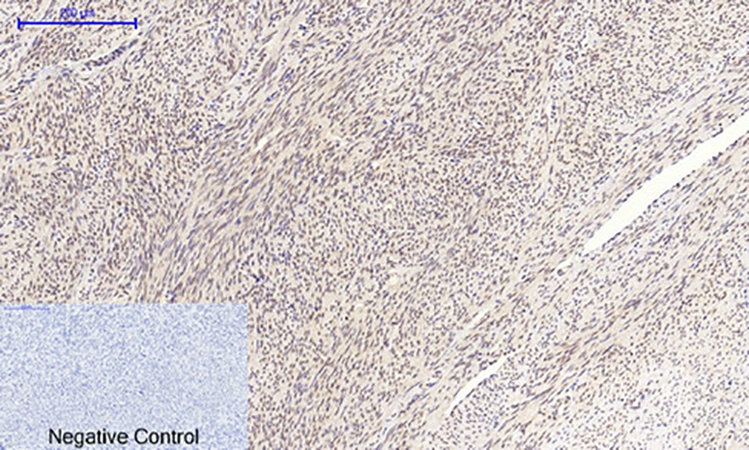
Fig.4. Immunohistochemical analysis of paraffin-embedded human uterus tissue. 1, c-Myc Polyclonal Antibody was diluted at 1:200 (4°C, overnight). 2, Sodium citrate pH 6.0 was used for antibody retrieval (>98°C, 20min). 3, secondary antibody was diluted at 1:200 (room temperature, 30min). Negative control was used by secondary antibody only.
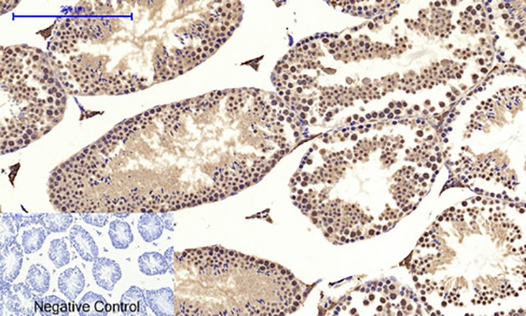
Fig.5. Immunohistochemical analysis of paraffin-embedded mouse testis tissue. 1, c-Myc Polyclonal Antibody was diluted at 1:200 (4°C, overnight). 2, Sodium citrate pH 6.0 was used for antibody retrieval (>98°C, 20min). 3, secondary antibody was diluted at 1:200 (room temperature, 30min). Negative control was used by secondary antibody only.
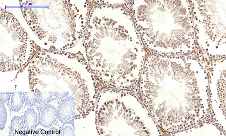
Fig.6. Immunohistochemical analysis of paraffin-embedded rat testis tissue. 1, c-Myc Polyclonal Antibody was diluted at 1:200 (4°C, overnight). 2, Sodium citrate pH 6.0 was used for antibody retrieval (>98°C, 20min). 3, secondary antibody was diluted at 1:200 (room temperature, 30min). Negative control was used by secondary antibody only.
You must be logged in to post a review.


Reviews
There are no reviews yet.