| Product name | Anti-GFP Tag Mouse Monoclonal Antibody (3D3) |
| Immunogen | Recombinant Protein |
| Host | Mouse |
| Reactivity | Mammals, Bacteria |
| Applications | IP, WB |
| Applications notes | Optimal working dilutions should be determined experimentally by the investigator. Suggested starting dilutions are as follows: WB (1:5000). |
| Clonality | Monoclonal |
| Preparation method | The antibody was affinity-purified from mouse ascites by affinity-chromatography using specific immunogen |
| Alternative | GFP; Green fluorescent protein |
| Formulation | Liquid solution |
| Storage buffer | Liquid in PBS, pH 7.4, containing 0.02% Sodium Azide as preservative and 50% Glycerol. |
| Storage instructions | Stable for one year at -20°C from date of shipment. For maximum recovery of product, centrifuge the original vial after thawing and prior to removing the cap. Aliquot to avoid repeated freezing and thawing. |
| Shipping | Gel pack with blue ice. |
| Precautions | The product listed herein is for research use only and is not intended for use in human or clinical diagnosis. Suggested applications of our products are not recommendations to use our products in violation of any patent or as a license. We cannot be responsible for patent infringements or other violations that may occur with the use of this product. |
| Background | The green fluorescent protein (GFP) is a protein composed of 238 amino acid residues (26.9kD) that exhibits bright green fluorescence when exposed to light in the blue to ultraviolet range. Although many other marine organisms have similar green fluorescent proteins, GFP traditionally refers to the protein first isolated from the jellyfish. The GFP has a major excitation peak at a wavelength of 395 nm and a minor one at 475 nm. Its emission peak is at 509 nm, which is in the lower green portion of the visible spectrum. |
| Alternative | GFP; Green fluorescent protein |
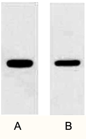
Fig. WB (1:10000) analysis of GFP fusion protein with Anti-GFP Mouse Monoclonal Antibody (3D3) in 1:5000 dilutions. 2ug GFP fusion protein(lane A) and 1 ug fusion protein(lane B).
Author:Meng, Li, et al. Publication name:Nature Communications IF:15.7
Author:Wang, Tao, et al Publication name:International Journal of Molecular Sciences IF:6
Author:Xia, Tian-Jin, et al. Publication name:BMC biology IF:4.5
Author:Songwattana, Pongpan, et al. Publication name: iScience IF:4.1
Author:L Yuan, Y Wang, BA Margulis, T Song, Z Wang, ... Publication name:Clinical Implant Dentistry and Related Research IF:0.8
You must be logged in to post a review.


Reviews
There are no reviews yet.