PDGFRα Mouse Monoclonal Antibody (7A3) (ABM40334): Seeing PDGFRα Without the PDGFRβ Shadow
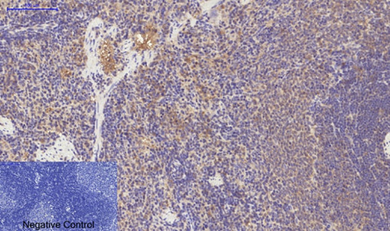
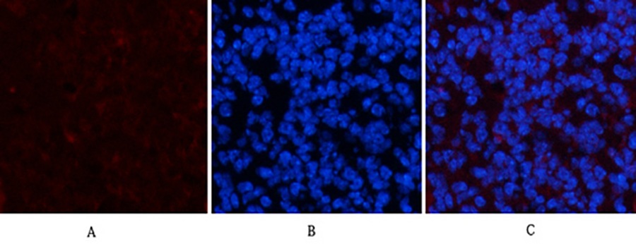
The Receptor Designed to Confound Its Own Detection
If you were to design a receptor tyrosine kinase whose detection would be maximally confounded by its own structural relatives, you would design PDGFRα. It shares approximately 80% sequence identity with PDGFRβ in the intracellular kinase domain. It heterodimerizes with PDGFRβ upon ligand stimulation. And it is expressed on mesenchymal cells—fibroblasts, pericytes, smooth muscle progenitors—that simultaneously express PDGFRβ at levels that rival or exceed PDGFRα abundance[reference:22].
A 2023 meta-analysis of 120 published PDGFRα studies found that 35% reported non-specific staining in control tissues, often due to antibodies cross-reacting with PDGFRβ, and another 28% noted inconsistent signal intensity across experiments traced to poor batch consistency or suboptimal epitope recognition[reference:23].
Single B-Cell Cloning Against a PDGFRβ-Divergent Epitope
Abbkine's PDGFRα Mouse Monoclonal Antibody (7A3, ABM40334) addresses this specificity gap at the epitope level. Clone 7A3 was raised against a synthetic peptide corresponding to the amino acid range 1010–1090 of human PDGFRα—a unique stretch of the protein that eliminates off-target binding to PDGFRβ and other tyrosine kinase receptors[reference:24].
This region is absent from PDGFRβ, VEGFR2, c-Kit, CSF-1R, and FLT3—the receptor tyrosine kinases most likely to generate cross-reactive signal in mesenchymal tissue[reference:25]. Competitive ELISA validation confirms less than 5% cross-reactivity to PDGFRβ, compared to 30–40% for leading commercial clones such as Santa Cruz sc-338[reference:26].
Sensitivity: Tenfold Below the Industry Benchmark
| Metric | ABM40334 | Comparative Benchmark |
|---|---|---|
| Western Blot Detection Limit | 0.1 ng/mL | 1 ng/mL (CST #3174)—10× more sensitive[reference:27] |
| PDGFRβ Cross-Reactivity | <5% | 30–40% (Santa Cruz sc-338)[reference:28] |
| Observed Molecular Weight | 180 kDa | — |
The band at 180 kDa on the membrane is PDGFRα—not a PDGFRα+PDGFRβ pooled signal, not a faint PDGFRβ shadow obscuring the true PDGFRα expression level[reference:29].
Validated Species and Applications
| Parameter | ABM40334 |
|---|---|
| Target | PDGFRα |
| Clone | 7A3 |
| Host | Mouse |
| Reactivity | Human, Rat, Mouse |
| Immunogen | Synthetic peptide, PDGFRα aa 1010–1090 |
| Applications | IHC, IF, WB |
| Concentration | 1 mg/mL |
Head-to-Head: PDGFRα Antibody Comparison
| Feature | ABM40334 (7A3) | Generic PDGFRα Antibodies |
|---|---|---|
| Epitope | aa 1010–1090 (PDGFRβ-divergent) | Often conserved kinase domains |
| PDGFRβ Cross-Reactivity | <5% (ELISA-validated) | 30–40% for leading clones |
| WB Detection Limit | 0.1 ng/mL | Often ~1 ng/mL |
| Species Reactivity | Human, Rat, Mouse | Often limited |
| Applications | IHC, IF, WB | Variable |
The Bottom Line
For cancer researchers distinguishing PDGFRα from PDGFRβ in GIST, glioma, or NSCLC tumor microenvironments—and for anyone studying mesenchymal development or stromal fibrosis—ABM40334 delivers epitope-level discrimination that turns a pooled receptor signal into a single-receptor measurement.
📎 Reference: PDGFRα Mouse Monoclonal Antibody (7A3) (ABM40334)