GFAP Monoclonal Antibody (ABM0021): Zero Cross-Reactivity in a Field Built on It
GFAP: The Astrocyte Marker That Consumed More Troubleshooting Hours Than Any Other
Glial fibrillary acidic protein (GFAP) is the defining marker of mature astrocytes—the class-III intermediate filament that assembles into 10-nm networks spanning the astrocyte cytoskeleton and distinguishes astrocytes from neurons, oligodendrocytes, microglia, and vascular cells in every brain section ever cut[reference:9].
It is also one of the most difficult proteins to detect without generating cross-reactive signal from its own intermediate filament relatives. Vimentin shares a class-III coiled-coil architecture with GFAP. Nestin shares an evolutionary origin. Desmin, peripherin, and internexin all populate the same molecular weight range and the same filamentous distribution[reference:10].
A polyclonal anti-GFAP antibody raised against full-length recombinant protein contains a subpopulation of immunoglobulins that bind these homologs. The resulting confocal image may look convincing—but the quantification is wrong, and the error propagates through every figure that depends on GFAP normalization[reference:11].
Single B-Cell Cloning Against a Divergent Rod-Domain Epitope
Abbkine's GFAP Monoclonal Antibody (ABM0021) was engineered via single B-cell cloning to target a conserved GFAP epitope spanning amino acids 150–200 of human GFAP—a region within the central rod domain where GFAP diverges from vimentin, nestin, and desmin at the primary sequence level[reference:12].
Unlike polyclonal alternatives whose epitope repertoire shifts from animal to animal and bleed to bleed, ABM0021 is a monoclonal immunoglobulin of defined specificity, affinity-purified from mouse ascites by affinity chromatography using the specific immunogen[reference:13].
The Performance Gap, in Numbers
| Metric | ABM0021 | Comparative Benchmark |
|---|---|---|
| Western Blot Detection Limit | 0.1 ng/mL | 1 ng/mL (CST #3655)—10× more sensitive[reference:14] |
| Inter-Assay CV | <3% | 15% (Abcam ab7260)—5× tighter[reference:15] |
| Cross-Reactivity (vimentin/nestin/desmin) | None observed | Variable with polyclonals |
That tenfold sensitivity increase allows detection of GFAP in dilute cerebrospinal fluid samples and microdissected astrocyte end-foot preparations where conventional antibodies produce blank lanes[reference:16].
Validated Applications and Protocols
| Application | Starting Dilution | Key Protocol Notes |
|---|---|---|
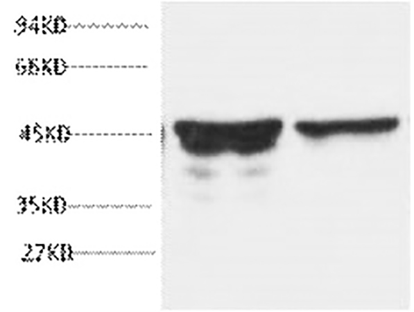
| Western Blot | 1:2000–1:5000 | Rat brain tissue at 1:5000 yields sharp 45 kDa band; 10% SDS-PAGE, PVDF transfer[reference:17] |
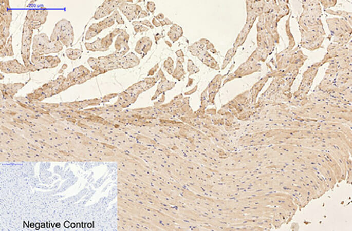
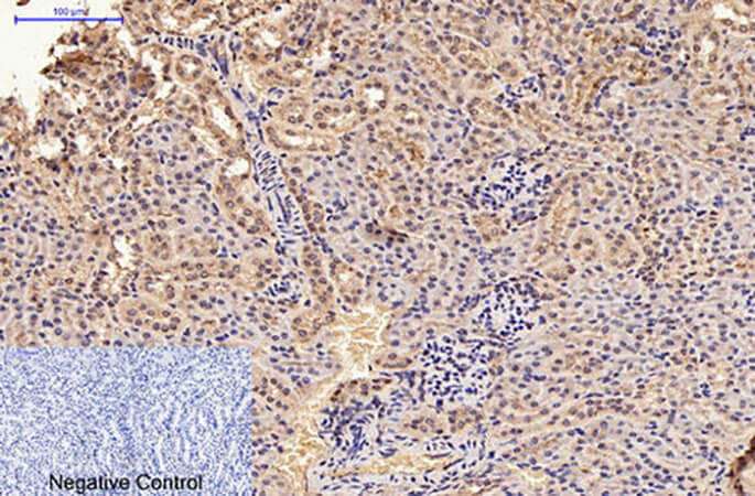
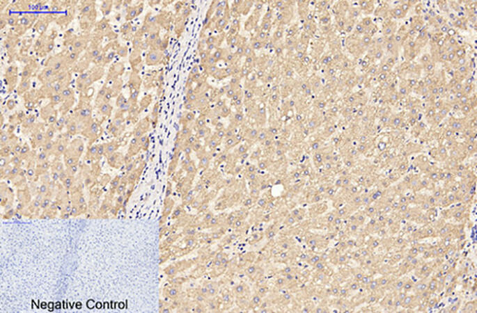
| IHC-P | 1:50–1:300 (1:200 optimal) | Sodium citrate buffer pH 6.0, >98°C 20 min; avoid harsh buffers that damage GFAP epitopes[reference:18] |
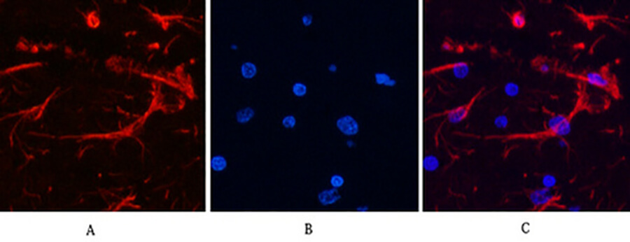
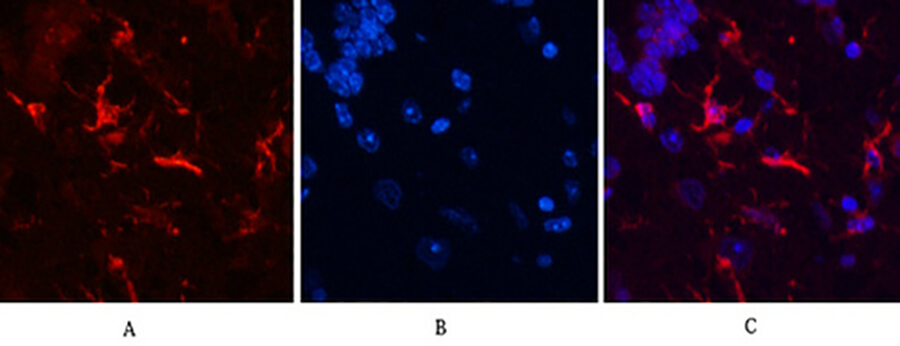
| Immunofluorescence | 1:200 | 4% PFA fixation 15 min; 0.1% Triton X-100 permeabilization[reference:19] |
The antibody detects all endogenous GFAP isoforms, critical for studying isoform switching in CNS disease[reference:20]. Validated for human, mouse, and rat tissues, including peripheral organs[reference:21].
Head-to-Head: GFAP Antibody Comparison
| Feature | ABM0021 | Polyclonal Anti-GFAP |
|---|---|---|
| Clonality | Monoclonal (single B-cell) | Polyclonal |
| Epitope | aa 150–200 (GFAP-divergent rod domain) | Multiple, including conserved regions |
| Cross-Reactivity | Zero to vimentin/nestin/desmin | Variable subpopulation binding homologs |
| WB Detection Limit | 0.1 ng/mL | Often ~1 ng/mL |
| Inter-Assay CV | <3% | Often 10–15% |
| Applications | WB, IHC, IF | Variable |
The Bottom Line
If you have ever squinted at a GFAP immunostain and wondered whether the filamentous signal in the corner of the image is an astrocyte or a vimentin-positive fibroblast, ABM0021 answers that question. It is the monoclonal that refuses to blur.
📎 Reference: GFAP Monoclonal Antibody (ABM0021)