| Product name | PTEN Polyclonal Antibody |
| Immunogen | Synthesized peptide derived from PTEN at AA range: 251-300 |
| Host | Rabbit |
| Reactivity | Human,Mouse,Rat |
| Applications | IF,WB,IHC,ELISA |
| Applications notes | Optimal working dilutions should be determined experimentally by the investigator. Suggested starting dilutions are as follows: IF 1:50-200;IHC: 100-300;WB 1:500-2000;ELISA 1:10000-20000 |
| Clonality | Polyclonal |
| Preparation method | The antibody was affinity-purified from rabbit antiserum by affinity-chromatography using epitope-specific immunogen. |
| Alternative | phosphatase and tensin homolog; phosphatase and tensin homolog pseudogene 1 |
| Formulation | Liquid solution |
| Concentration | 1 mg/ml |
| Molecular weight | 50kD |
| Storage buffer | Liquid in PBS containing 50% glycerol, 0.5% BSA and 0.02% sodium azide. |
| Storage instructions | Stable for one year at -20°C from date of shipment. For maximum recovery of product, centrifuge the original vial after thawing and prior to removing the cap. Aliquot to avoid repeated freezing and thawing. |
| Shipping | Gel pack with blue ice. |
| Precautions | The product listed herein is for research use only and is not intended for use in human or clinical diagnosis. Suggested applications of our products are not recommendations to use our products in violation of any patent or as a license. We cannot be responsible for patent infringements or other violations that may occur with the use of this product. |
| Background | PTEN (phosphatase and tensin homolog) was identified as a tumor suppressor that is mutated in a large number of cancers at high frequency. The protein encoded by PTEN is a phosphatidylinositol-3,4,5-trisphosphate 3-phosphatase. It contains a tensin like domain as well as a catalytic domain similar to that of the dual specificity protein tyrosine phosphatases. Unlike most of the protein tyrosine phosphatases, this protein preferentially dephosphorylates phosphoinositide substrates. It negatively regulates intracellular levels of phosphatidylinositol-3,4,5-trisphosphate in cells and functions as a tumor suppressor by negatively regulating AKT/PKB signaling pathway. The use of a non-canonical (CUG) upstream initiation site produces a longer isoform that initiates translation with a leucine, and is thought to be preferentially associated with the mitochondrial inner membrane. This longer isoform may help regulate energy metabolism in the mitochondria. A pseudogene of PTEN is found on chromosome 9. Alternative splicing and the use of multiple translation start codons results in multiple transcript variants encoding different isoforms. |
| Gene ID | 5728 |
| Alternative | phosphatase and tensin homolog; phosphatase and tensin homolog pseudogene 1 |
| Others | PTEN Polyclonal Antibody detects endogenous levels of PTEN. |
| Accession | P60484 |
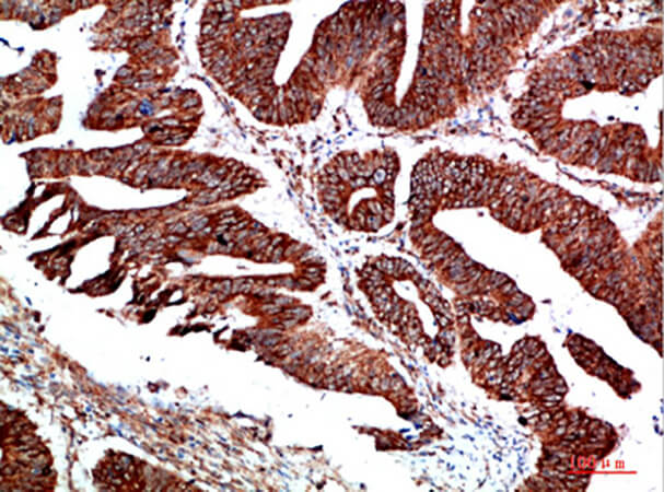
Fig.1. Immunohistochemical analysis of paraffin-embedded human colon cancer, antibody was diluted at 1:200.
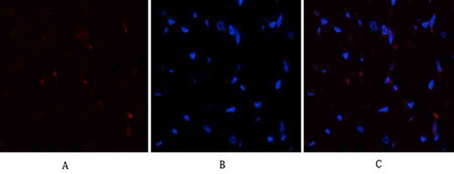
Fig.2. Immunofluorescence analysis of human-heart tissue. 1, PTEN Polyclonal Antibody (red) was diluted at 1:200 (4°C, overnight). 2, Cy3 Labeled secondary antibody was diluted at 1:300 (room temperature, 50min). 3, Picture B: DAPI (blue) 10min. Picture A: Target. Picture B: DAPI. Picture C: merge of A+B.
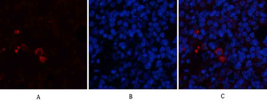
Fig.3. Immunofluorescence analysis of rat spleen tissue. 1, PTEN Polyclonal Antibody (red) was diluted at 1:200 (4°C, overnight). 2, Cy3 Labeled secondary antibody was diluted at 1:300 (room temperature, 50min). 3, Picture B: DAPI (blue) 10min. Picture A: Target. Picture B: DAPI. Picture C: merge of A+B.
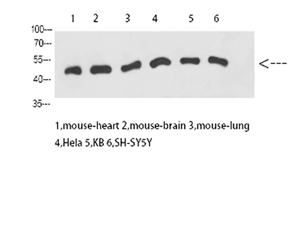
Fig.4. Western Blot analysis of mouse heart (1), mouse brain (2), mouse lung (3), Hela (4), KB (5), SH-SY5Y (6), diluted at 1:1000.
You must be logged in to post a review.


Reviews
There are no reviews yet.