| Product name | CD63 Polyclonal Antibody |
| Immunogen | Synthesized peptide derived from the Internal region of human CD63 |
| Host | Rabbit |
| Reactivity | Human,Rat,Mouse, |
| Applications | IF,WB,IHC,ELISA |
| Applications notes | Optimal working dilutions should be determined experimentally by the investigator. Suggested starting dilutions are as follows: IF 1:50-200;WB 1:500-1:2000;IHC: 1:100-1:300;ELISA 1:20000;Not yet tested in other applications; |
| Clonality | Polyclonal |
| Preparation method | The antibody was affinity-purified from rabbit antiserum by affinity-chromatography using epitope-specific immunogen. |
| Alternative | CD63; MLA1; TSPAN30; CD63 antigen; Granulophysin; Lysosomal-associated membrane protein 3; LAMP-3; Melanoma-associated antigen ME491; OMA81H; Ocular melanoma-associated antigen; Tetraspanin-30; Tspan-30; CD63 |
| Formulation | Liquid solution |
| Concentration | 1 mg/ml |
| Molecular weight | 26,35-65(kD |
| Storage buffer | Liquid in PBS containing 50% glycerol, 0.5% BSA and 0.02% sodium azide. |
| Storage instructions | Stable for one year at -20°C from date of shipment. For maximum recovery of product, centrifuge the original vial after thawing and prior to removing the cap. Aliquot to avoid repeated freezing and thawing. |
| Shipping | Gel pack with blue ice. |
| Precautions | The product listed herein is for research use only and is not intended for use in human or clinical diagnosis. Suggested applications of our products are not recommendations to use our products in violation of any patent or as a license. We cannot be responsible for patent infringements or other violations that may occur with the use of this product. |
| Background | CD63 molecule encoded by CD63 is a member of the transmembrane 4 superfamily, also known as the tetraspanin family. Most of these members are cell-surface proteins that are characterized by the presence of four hydrophobic domains. The proteins mediate signal transduction events that play a role in the regulation of cell development, activation, growth and motility. The encoded protein is a cell surface glycoprotein that is known to complex with integrins. It may function as a blood platelet activation marker. Deficiency of this protein is associated with Hermansky-Pudlak syndrome. Also this gene has been associated with tumor progression. Alternative splicing results in multiple transcript variants encoding different protein isoforms. |
| Gene ID | 967 |
| Alternative | CD63; MLA1; TSPAN30; CD63 antigen; Granulophysin; Lysosomal-associated membrane protein 3; LAMP-3; Melanoma-associated antigen ME491; OMA81H; Ocular melanoma-associated antigen; Tetraspanin-30; Tspan-30; CD63 |
| Others | CD63 Polyclonal Antibody detects endogenous levels of CD63 protein. |
| Accession | P08962 |
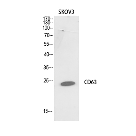
Fig.1. Western Blot analysis of SKOV3 cells using CD63 Polyclonal Antibody.
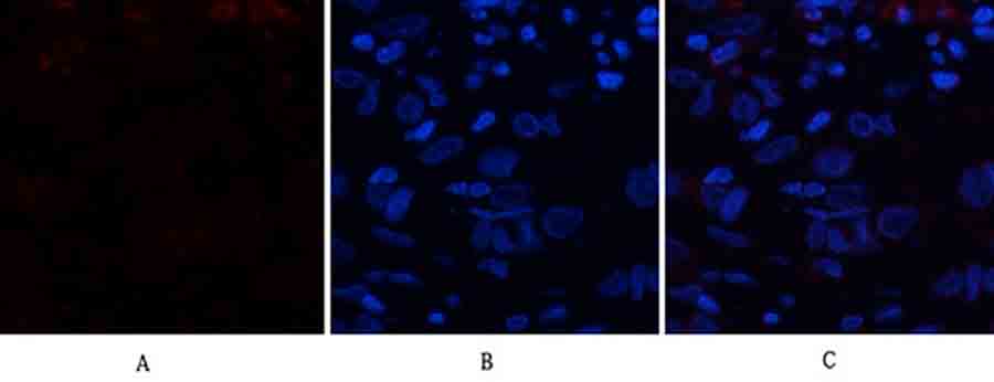
Fig.2. Immunofluorescence analysis of human breast cancer tissue. 1, CD63 Polyclonal Antibody (red) was diluted at 1:200 (4°C, overnight). 2, Cy3 Labeled secondary antibody was diluted at 1:300 (room temperature, 50min). 3, Picture B: DAPI (blue) 10min. Picture A: Target. Picture B: DAPI. Picture C: merge of A+B.
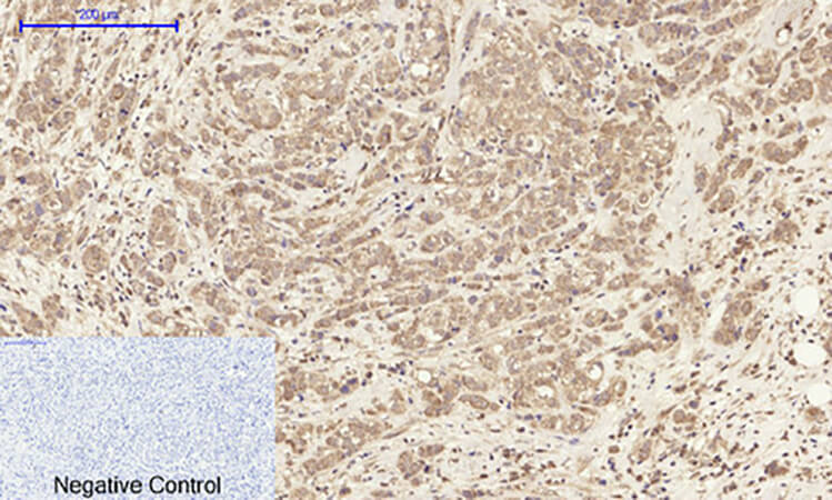
Fig.3. Immunohistochemical analysis of paraffin-embedded human breast cancer tissue. 1, CD63 Polyclonal Antibody was diluted at 1:200 (4°C, overnight). 2, Sodium citrate pH 6.0 was used for antibody retrieval (>98°C, 20min). 3, secondary antibody was diluted at 1:200 (room temperature, 30min). Negative control was used by secondary antibody only.
You must be logged in to post a review.


Reviews
There are no reviews yet.