| Product name | Anti-PCNA Mouse Monoclonal Antibody (1D7) |
| Immunogen | Synthetic Peptide |
| Host | Mouse |
| Reactivity | Human, Mouse, Rat |
| Applications | IF, IHC-P, WB |
| Applications notes | Optimal working dilutions should be determined experimentally by the investigator. Suggested starting dilutions are as follows: WB (1:5000), IHC-P (1:200), IF (1:200). |
| Clonality | Monoclonal |
| Preparation method | The antibody was affinity-purified from mouse ascites by affinity-chromatography using specific immunogen |
| Alternative | PCNA; Proliferating cell nuclear antigen; PCNA; Cyclin |
| Formulation | Liquid solution |
| Storage buffer | Liquid in PBS, pH 7.4, containing 0.02% Sodium Azide as preservative and 50% Glycerol. |
| Storage instructions | Stable for one year at -20°C from date of shipment. For maximum recovery of product, centrifuge the original vial after thawing and prior to removing the cap. Aliquot to avoid repeated freezing and thawing. |
| Shipping | Gel pack with blue ice. |
| Precautions | The product listed herein is for research use only and is not intended for use in human or clinical diagnosis. Suggested applications of our products are not recommendations to use our products in violation of any patent or as a license. We cannot be responsible for patent infringements or other violations that may occur with the use of this product. |
| Background | Proliferating Cell Nuclear Antigen, commonly known as PCNA, is a protein that acts as a processivity factor for DNA polymerase in eukaryotic cells. It achieves this processivity by encircling the DNA, thus creating a topological link to the genome. PCNA was originally identified as an antigen that is expressed in the nuclei of cells during the DNA synthesis phase of the cell cycle. |
| Gene ID | 5111 |
| Alternative | PCNA; Proliferating cell nuclear antigen; PCNA; Cyclin |
| Accession | P12004 |
| Observed Band(KD) | 29 |
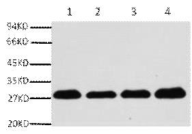
Fig.1. Western blot analysis of Hela (1), rat brain (2), NIH 3T3(3), 293T(4), diluted at 1:5000.
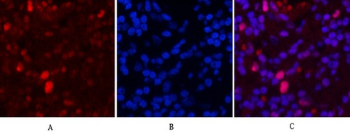
Fig.2. Immunofluorescence analysis of human lung cancer tissue. 1, PCNA Monoclonal Antibody (1D7) (red) was diluted at 1:200 (4°C, overnight). 2, Cy3 Labeled secondary antibody was diluted at 1:300 (room temperature, 50min). 3, Picture B: DAPI (blue) 10min. Picture A: Target. Picture B: DAPI. Picture C: merge of A+B.
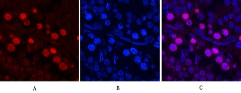
Fig.3. Immunofluorescence analysis of rat testis tissue. 1, PCNA Monoclonal Antibody (1D7) (red) was diluted at 1:200 (4°C, overnight). 2, Cy3 Labeled secondary antibody was diluted at 1:300 (room temperature, 50min). 3, Picture B: DAPI (blue) 10min. Picture A: Target. Picture B: DAPI. Picture C: merge of A+B.
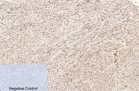
Fig.4. Immunohistochemical analysis of paraffin-embedded human uterus tissue. 1, PCNA Monoclonal Antibody (1D7) was diluted at 1:200 (4°C, overnight). 2, Sodium citrate pH 6.0 was used for antibody retrieval(>98°C, 20min). 3, secondary antibody was diluted at 1:200 (room temperature, 30min). Negative control was used by secondary antibody only.
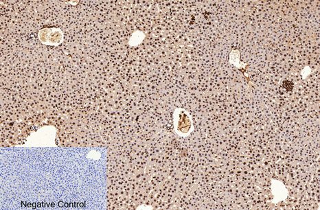
Fig.5. Immunohistochemical analysis of paraffin-embedded mouse liver tissue. 1, PCNA Monoclonal Antibody (1D7) was diluted at 1:200 (4°C, overnight). 2, Sodium citrate pH 6.0 was used for antibody retrieval(>98°C, 20min). 3, secondary antibody was diluted at 1:200 (room temperature, 30min). Negative control was used by secondary antibody only.
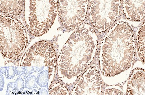
Fig.6. Immunohistochemical analysis of paraffin-embedded rat testis tissue. 1, PCNA Monoclonal Antibod y(1D7) was diluted at 1:200 (4°C, overnight). 2, Sodium citrate pH 6.0 was used for antibody retrieval(>98°C, 20min). 3, secondary antibody was diluted at 1:200 (room temperature, 30min). Negative control was used by secondary antibody only.
Author:Xia M M, Li Z, Chen Y Y, et al Publication name:Sensors and Actuators B: Chemical IF:6
Author:Wang, Hong-Jin, et al. Publication name:Clinical, Cosmetic and Investigational Dermatology IF:4
Author:Feng, Mao, et al Publication name:Journal of proteome research 14 IF:4
Author:Xu X H, Ding D F, Yong H J, et al Publication name:EUR REV MED PHARMACO IF:2
Author:Cai R, Jiang Y, Yang W, et al Publication name:Journal of microbiology and biotechnology IF:2
You must be logged in to post a review.


Reviews
There are no reviews yet.