| Product name | Anti-COX IV Mouse Monoclonal Antibody (14Y2) |
| Immunogen | Recombinant Protein |
| Host | Mouse |
| Reactivity | Human, Mouse, Rat |
| Applications | IF, IHC, WB |
| Applications notes | Optimal working dilutions should be determined experimentally by the investigator. Suggested starting dilutions are as follows: WB (1:1000-1:3000), IHC-P (1:50-1:300), IF (1:200). |
| Clonality | Monoclonal |
| Preparation method | The antibody was affinity-purified from mouse ascites by affinity-chromatography using specific immunogen |
| Alternative | COX4I1; COX4; Cytochrome c oxidase subunit 4 isoform 1; mitochondrial; Cytochrome c oxidase polypeptide IV; Cytochrome c oxidase subunit IV isoform 1; COX IV-1 |
| Formulation | Liquid solution |
| Storage buffer | Liquid in PBS, pH 7.4, containing 0.02% Sodium Azide as preservative and 50% Glycerol. |
| Storage instructions | Stable for one year at -20°C from date of shipment. For maximum recovery of product, centrifuge the original vial after thawing and prior to removing the cap. Aliquot to avoid repeated freezing and thawing. |
| Shipping | Gel pack with blue ice. |
| Precautions | The product listed herein is for research use only and is not intended for use in human or clinical diagnosis. Suggested applications of our products are not recommendations to use our products in violation of any patent or as a license. We cannot be responsible for patent infringements or other violations that may occur with the use of this product. |
| Background | Cytochrome c Oxidase or Complex IV (EC 1.9.3.1) is a large transmembrane protein complex found in bacteria and the mitochondrion. It is the last enzyme in the respiratory electron transport chain of mitochondria (or bacteria) located in the mitochondrial (or bacterial) membrane. It receives an electron from each of four cytochrome c molecules, and transfers them to one oxygen molecule, converting molecular oxygen to two molecules of water. |
| Gene ID | 1327 |
| Alternative | COX4I1; COX4; Cytochrome c oxidase subunit 4 isoform 1; mitochondrial; Cytochrome c oxidase polypeptide IV; Cytochrome c oxidase subunit IV isoform 1; COX IV-1 |
| Accession | P13073 |
| Observed Band(KD) | 15 |
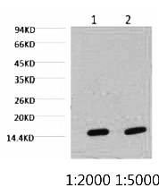
Fig.1. Western blot analysis of Hela, diluted at 1) 1:2000 2) 1:5000.
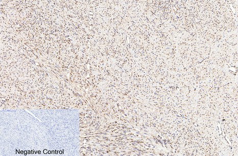
Fig.2. Immunohistochemical analysis of paraffin-embedded human uterus cancer tissue. 1, COX IV Monoclonal Antibody (14Y2) was diluted at 1:200 (4°C, overnight). 2, Sodium citrate pH 6.0 was used for antibody retrieval (>98°C, 20min). 3, secondary antibody was diluted at 1:200 (room temperature, 30min). Negative control was used by secondary antibody only.
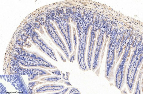
Fig.3. Immunohistochemical analysis of paraffin-embedded mouse colon tissue. 1, COX IV Monoclonal Antibody (14Y2) was diluted at 1:200 (4°C, overnight). 2, Sodium citrate pH 6.0 was used for antibody retrieval (>98°C, 20min). 3, secondary antibody was diluted at 1:200 (room temperature, 30min). Negative control was used by secondary antibody only.
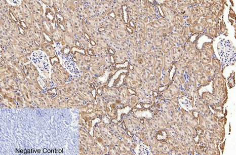
Fig.4. Immunohistochemical analysis of paraffin-embedded rat kidney tissue. 1, COX IV Monoclonal Antibody (14Y2) was diluted at 1:200 (4°C, overnight). 2, Sodium citrate pH 6.0 was used for antibody retrieval (>98°C, 20min). 3, secondary antibody was diluted at 1:200 (room temperature, 30min). Negative control was used by secondary antibody only.
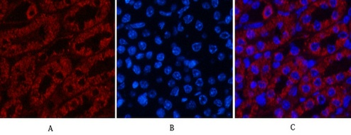
Fig.5. Immunofluorescence analysis of mouse kidney tissue. 1, COX IV Monoclonal Antibody (14Y2) (red) was diluted at 1:200 (4°C, overnight). 2, Cy3 Labeled secondary antibody was diluted at 1:300 (room temperature, 50min). 3, Picture B: DAPI (blue) 10min. Picture A: Target. Picture B: DAPI. Picture C: merge of A+B.
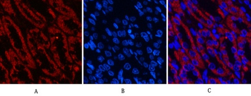
Fig.6. Immunofluorescence analysis of rat kidney tissue. 1, COX IV Monoclonal Antibody (14Y2) (red) was diluted at 1:200 (4°C, overnight). 2, Cy3 Labeled secondary antibody was diluted at 1:300 (room temperature, 50min). 3, Picture B: DAPI (blue) 10min. Picture A: Target. Picture B: DAPI. Picture C: merge of A+B.
Author:T Wang, L Mo, J Ou, Q Fang, H Wu, Y Wu Publication name:Frontiers in immunology IF:8
Author:Tao L, Qiu Y, Fu X, et al Publication name:Journal of neuroinflammation IF:6
Author:Zhang Y Z, An J H, Liu Y, et al Publication name:Cell Physiol Biochem IF:5
Author:Zhang Y, Zhang C, Li J, Jiang M, Guo S, Yang G, Zhang L, Wang F, Yi S, Wang J, Fu Y, Zhang Y Publication name:Cell Commun Signal IF:4
Author:Chen, Xin, et al Publication name:Brain Research 1625 (2015): 275-286 IF:3
Author:Shao L, Chian R C, Xu Y, et al Publication name:Reproduction, Fertility and Development IF:2
Author:Tian C H, Xie X M, Li Y, et al Publication name:China Biotechnology IF:/
You must be logged in to post a review.


Reviews
There are no reviews yet.