


ABP53233: Abbkine’s HMG-1 Polyclonal Antibody – A Versatile Tool for HMGB1 Research in Inflammation, Cancer, and Nuclear Biology
For researchers delving into nuclear biology, inflammation, or cancer metastasis, High Mobility Group Box 1 (HMG-1, also known as HMGB1) stands as a pivotal target—yet detecting this multifunctional protein with precision has long been a bottleneck. As a non-histone nuclear protein, HMGB1 regulates transcription and DNA organization in the nucleus, while its extracellular release drives proinflammatory responses and tumor cell migration, linking it to diseases from sepsis to solid tumors. To address the need for a reliable, specific reagent, Abbkine has developed the HMG-1 Polyclonal Antibody (catalog number: ABP53233), priced at $109 for 30μl. This antibody isn’t just a detection tool; it’s a tailored solution for decoding HMGB1’s complex roles across biological contexts, combining high specificity, cross-species compatibility, and multi-application flexibility that aligns with cutting-edge research demands.
What sets Abbkine’s ABP53233 apart lies in its targeted immunogen design— a synthesized peptide derived from the N-terminal region of human HMG-1. This choice is no accident: the N-terminus of HMGB1 contains key DNA-binding domains and is critical for its nuclear function and extracellular proinflammatory activity. By focusing on this region, the antibody ensures specificity for endogenous HMG-1 (HMGB1) protein, avoiding cross-reactivity with other HMG family members or unrelated proteins—a common pitfall in HMGB1 research. Produced in rabbit, this polyclonal antibody offers an additional edge: recognition of multiple epitopes on the HMG-1 protein, which enhances signal robustness, even when detecting low-abundance or post-translationally modified HMGB1 (e.g., acetylated or phosphorylated forms involved in nuclear export). Its reactivity with human, mouse, and rat samples further extends its utility, enabling seamless translation from preclinical animal models (e.g., mouse sepsis or rat tumor xenografts) to human tissue samples—eliminating the need for multiple antibodies and ensuring consistent data across experimental systems.
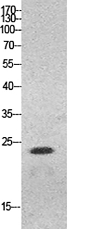
When it comes to application flexibility, this HMG-1 polyclonal antibody stands out as a multi-functional tool, validated for four core techniques: Immunofluorescence (IF), Western Blot (WB), Immunohistochemistry (IHC), and Enzyme-Linked Immunosorbent Assay (ELISA). Each application addresses distinct research questions, and ABP53233 delivers tailored performance for each. For WB, the antibody detects a clear band at ~25 kDa (consistent with the product’s observed band data), slightly lower than the theoretical 30 kDa—likely due to post-translational processing, a detail that reflects its ability to recognize native, functionally relevant HMGB1 forms. The suggested starting dilution of 1:500–2000 balances sensitivity and signal-to-noise ratio, making it ideal for quantifying HMGB1 expression in cell lysates (e.g., HeLa cells, as shown in product images). For IHC, it performs reliably in paraffin-embedded tissues from humans (uterus), mice (heart), and rats (lung) at 1:100–300 dilution, with clear staining and minimal background (supported by negative control data)—a boon for studying HMGB1 distribution in pathological tissues like tumor biopsies or inflamed organs. IF (1:50–200 dilution) enables subcellular localization, critical for distinguishing nuclear HMGB1 (transcriptional regulation) from cytoplasmic/extracellular HMGB1 (inflammatory signaling). Finally, its high sensitivity for ELISA (1:20000 dilution) allows quantitative measurement of HMGB1 in biological fluids (e.g., serum, cell supernatants), essential for studies on cytokine release in sepsis or tumor microenvironments.
Practicality in lab workflows is often the unsung hero of reliable research reagents, and ABP53233 excels here with design choices that reduce friction and enhance reproducibility. Supplied as a ready-to-use liquid solution at 1 mg/ml, it eliminates the need for reconstitution—saving time and avoiding concentration errors common with lyophilized antibodies. The storage buffer (PBS with 50% glycerol, 0.5% BSA, and 0.02% sodium azide) preserves activity for up to one year at -20°C, while the recommendation to centrifuge after thawing and aliquot to avoid repeated freeze-thaw cycles is a pragmatic touch that extends the antibody’s lifespan. Shipping with gel packs and blue ice ensures temperature stability during transit, a critical factor for antibody performance that many budget reagents overlook. For labs prioritizing consistency and efficiency, these features translate to fewer failed experiments and more reliable data—especially important when working with a protein as dynamically regulated as HMGB1.
To truly appreciate the value of ABP53233, we must connect its performance to the biological significance of HMGB1. Encoded by the HMGB1 gene (Gene ID: 3146), this protein is a “double-edged sword”: in the nucleus, it maintains chromatin structure and regulates gene expression; extracellularly, it acts as a damage-associated molecular pattern (DAMP) that triggers immune responses. Dysregulation of HMGB1 is linked to a wide range of diseases—sepsis (where its release drives cytokine storms), cancer (promoting tumor cell migration and angiogenesis), and autoimmune disorders (exacerbating inflammation). As research into HMGB1-targeted therapies accelerates (e.g., neutralizing antibodies for sepsis or cancer), the need for precise detection tools becomes urgent. ABP53233’s ability to detect endogenous HMGB1 across species and applications makes it invaluable for preclinical studies: for example, validating HMGB1 expression in tumor models before testing inhibitors, or quantifying HMGB1 release in sepsis models to assess therapeutic efficacy. Unlike antibodies that only recognize recombinant HMGB1, ABP53233 mirrors physiological conditions, ensuring data that translates to clinical relevance.
An underdiscussed advantage of polyclonal antibodies for HMGB1 detection is their resilience to structural variations—and ABP53233 leverages this perfectly. HMGB1 undergoes conformational changes when released from the nucleus (e.g., disulfide bond reduction), which can mask epitopes recognized by monoclonals. As a polyclonal antibody, ABP53233 binds multiple epitopes, including those that remain accessible after structural changes, making it more reliable for detecting HMGB1 in complex biological samples (e.g., serum from sepsis patients or tumor tissue lysates). Additionally, Abbkine’s affinity-purification process—using the epitope-specific N-terminal immunogen—ensures high purity, reducing non-specific binding and background noise. This combination of resilience and purity is particularly valuable for studies where HMGB1’s structural state is a key variable, a nuance that many generic antibodies fail to address.
For researchers aiming to unlock HMGB1’s roles in disease or develop targeted therapies, Abbkine’s ABP53233 HMG-1 Polyclonal Antibody is a standout choice—marrying specificity, versatility, and practicality with alignment to high-impact research areas. Whether you’re quantifying HMGB1 in Western blots, localizing it in tissue sections via IHC/IF, or measuring its levels in biological fluids with ELISA, this antibody delivers consistent, reliable results across human, mouse, and rat samples. To explore detailed application protocols, access product images, or secure your vial, visit the official product page: https://www.abbkine.com/product/hmg-1-polyclonal-antibody-abp53233/.


