| Product name | CK16 Monoclonal Antibody |
| Immunogen | Synthetic Peptide |
| Host | Mouse |
| Reactivity | Human,Mouse,Rat |
| Applications | IHC,IF |
| Applications notes | Optimal working dilutions should be determined experimentally by the investigator. Suggested starting dilutions are as follows: IHC 1:50-200;IF 1:50-200 |
| Clonality | Monoclonal |
| Preparation method | The antibody was affinity-purified from mouse ascites by affinity-chromatography using specific immunogen. |
| Alternative | KRT16; KRT16A; Keratin, type I cytoskeletal 16; Cytokeratin-16; CK-16; Keratin-16; K16 |
| Formulation | Liquid solution |
| Concentration | 1 mg/ml |
| Molecular weight | 51kD |
| Storage buffer | PBS, pH 7.4, containing 0.5%BSA, 0.02% sodium azide as Preservative and 50% Glycerol. |
| Storage instructions | Stable for one year at -20°C from date of shipment. For maximum recovery of product, centrifuge the original vial after thawing and prior to removing the cap. Aliquot to avoid repeated freezing and thawing. |
| Shipping | Gel pack with blue ice. |
| Precautions | The product listed herein is for research use only and is not intended for use in human or clinical diagnosis. Suggested applications of our products are not recommendations to use our products in violation of any patent or as a license. We cannot be responsible for patent infringements or other violations that may occur with the use of this product. |
| Background | keratin 16 encoded by KRT16 is a member of the keratin gene family. The keratins are intermediate filament proteins responsible for the structural integrity of epithelial cells and are subdivided into cytokeratins and hair keratins. Most of the type I cytokeratins consist of acidic proteins which are arranged in pairs of heterotypic keratin chains and are clustered in a region of chromosome 17q12-q21. This keratin has been coexpressed with keratin 14 in a number of epithelial tissues, including esophagus, tongue, and hair follicles. Mutations in KRT16 are associated with type 1 pachyonychia congenita, non-epidermolytic palmoplantar keratoderma and unilateral palmoplantar verrucous nevus. |
| Gene ID | 3868 |
| Alternative | KRT16; KRT16A; Keratin, type I cytoskeletal 16; Cytokeratin-16; CK-16; Keratin-16; K16 |
| Others | The antibody detects endogenous CK16 proteins. |
| Accession | P08779 |
| Observed Band(KD) | 51 |
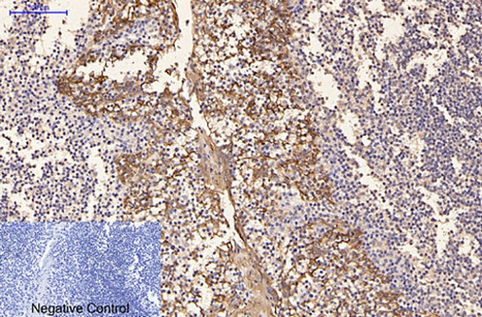
Fig.1. Immunohistochemical analysis of paraffin-embedded human tonsil tissue. 1, CK16 Monoclonal Antibody was diluted at 1:200 (4°C, overnight). 2, Sodium citrate pH 6.0 was used for antibody retrieval (>98°C, 20min). 3, secondary antibody was diluted at 1:200 (room temperature, 30min). Negative control was used by secondary antibody only.
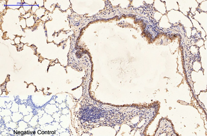
Fig.2. Immunohistochemical analysis of paraffin-embedded rat lung tissue. 1, CK16 Monoclonal Antibody was diluted at 1:200 (4°C, overnight). 2, Sodium citrate pH 6.0 was used for antibody retrieval (>98°C, 20min). 3, secondary antibody was diluted at 1:200 (room temperature, 30min). Negative control was used by secondary antibody only.
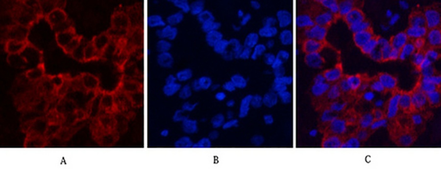
Fig.3. Immunofluorescence analysis of human liver cancer tissue. 1, CK16 Monoclonal Antibody (red) was diluted at 1:200 (4°C, overnight). 2, Cy3 Labeled secondary antibody was diluted at 1:300 (room temperature, 50min). 3, Picture B: DAPI (blue) 10min. Picture A: Target. Picture B: DAPI. Picture C: merge of A+B.
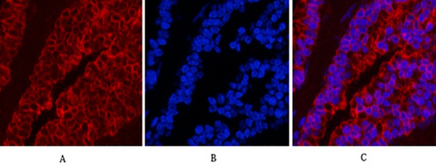
Fig.4. Immunofluorescence analysis of mouse lung tissue. 1, CK16 Monoclonal Antibody (red) was diluted at 1:200 (4°C, overnight). 2, Cy3 Labeled secondary antibody was diluted at 1:300 (room temperature, 50min). 3, Picture B: DAPI (blue) 10min. Picture A: Target. Picture B: DAPI. Picture C: merge of A+B.
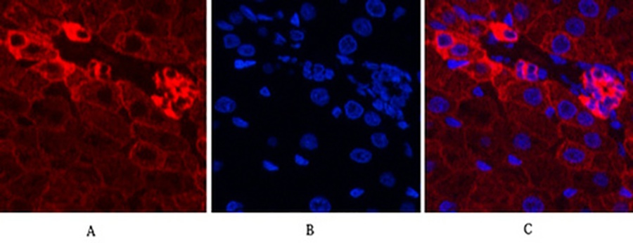
Fig.5. Immunofluorescence analysis of rat liver tissue. 1, CK16 Monoclonal Antibody (red) was diluted at 1:200 (4°C, overnight). 2, Cy3 Labeled secondary antibody was diluted at 1:300 (room temperature, 50min). 3, Picture B: DAPI (blue) 10min. Picture A: Target. Picture B: DAPI. Picture C: merge of A+B.
You must be logged in to post a review.


Reviews
There are no reviews yet.