| Product name | CD4 Monoclonal Antibody |
| Immunogen | Synthetic Peptide |
| Host | Mouse |
| Reactivity | Human,Mouse,Rat |
| Applications | IHC |
| Applications notes | Optimal working dilutions should be determined experimentally by the investigator. Suggested starting dilutions are as follows: IHC 1:200 |
| Clonality | Monoclonal |
| Preparation method | The antibody was affinity-purified from mouse ascites by affinity-chromatography using specific immunogen. |
| Alternative | CD4; T-cell surface glycoprotein CD4; T-cell surface antigen T4/Leu-3; CD4 |
| Formulation | Liquid solution |
| Concentration | 1 mg/ml |
| Storage buffer | PBS, pH 7.4, containing 0.5%BSA, 0.02% sodium azide as Preservative and 50% Glycerol. |
| Storage instructions | Stable for one year at -20°C from date of shipment. For maximum recovery of product, centrifuge the original vial after thawing and prior to removing the cap. Aliquot to avoid repeated freezing and thawing. |
| Shipping | Gel pack with blue ice. |
| Precautions | The product listed herein is for research use only and is not intended for use in human or clinical diagnosis. Suggested applications of our products are not recommendations to use our products in violation of any patent or as a license. We cannot be responsible for patent infringements or other violations that may occur with the use of this product. |
| Background | CD4 encodes a membrane glycoprotein of T lymphocytes that interacts with major histocompatibility complex class II antigenes and is also a receptor for the human immunodeficiency virus. CD4 is expressed not only in T lymphocytes, but also in B cells, macrophages, and granulocytes. It is also expressed in specific regions of the brain. CD4 molecule functions to initiate or augment the early phase of T-cell activation, and may function as an important mediator of indirect neuronal damage in infectious and immune-mediated diseases of the central nervous system. Multiple alternatively spliced transcript variants encoding different isoforms have been identified in CD4 . |
| Gene ID | 920 |
| Alternative | CD4; T-cell surface glycoprotein CD4; T-cell surface antigen T4/Leu-3; CD4 |
| Others | The antibody detects endogenous CD4 proteins. |
| Accession | P01730 |
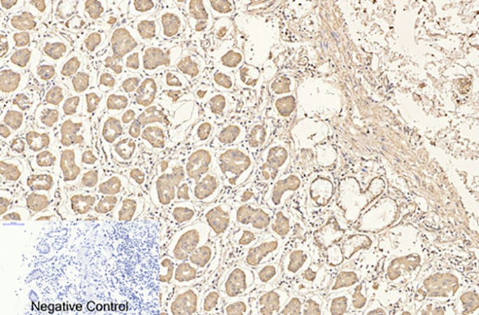
Fig.1. Immunohistochemical analysis of paraffin-embedded human stomach tissue. 1, CD4 Monoclonal Antibody was diluted at 1:200 (4°C, overnight). 2, Sodium citrate pH 6.0 was used for antibody retrieval (>98°C, 20min). 3, secondary antibody was diluted at 1:200 (room temperature, 30min). Negative control was used by secondary antibody only.
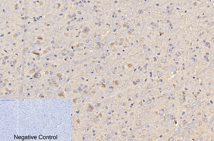
Fig.2. Immunohistochemical analysis of paraffin-embedded mouse brain tissue. 1, CD4 Monoclonal Antibody was diluted at 1:200 (4°C, overnight). 2, Sodium citrate pH 6.0 was used for antibody retrieval (>98°C, 20min). 3, secondary antibody was diluted at 1:200 (room temperature, 30min). Negative control was used by secondary antibody only.
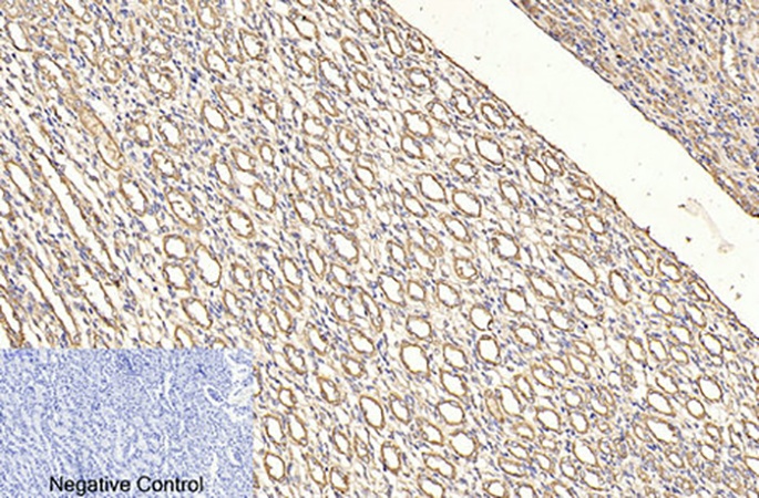
Fig.3. Immunohistochemical analysis of paraffin-embedded rat kidney tissue. 1, CD4 Monoclonal Antibody was diluted at 1:200 (4°C, overnight). 2, Sodium citrate pH 6.0 was used for antibody retrieval (>98°C, 20min). 3, secondary antibody was diluted at 1:200 (room temperature, 30min). Negative control was used by secondary antibody only.
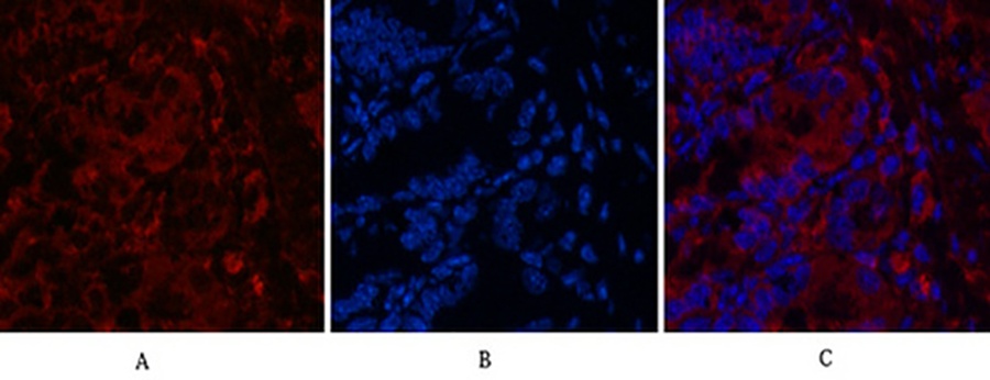
Fig.4. Immunofluorescence analysis of mouse colon tissue. 1, CD4 Monoclonal Antibody (red) was diluted at 1:200 (4°C, overnight). 2, Cy3 Labeled secondary antibody was diluted at 1:300 (room temperature, 50min). 3, Picture B: DAPI (blue) 10min. Picture A: Target. Picture B: DAPI. Picture C: merge of A+B.
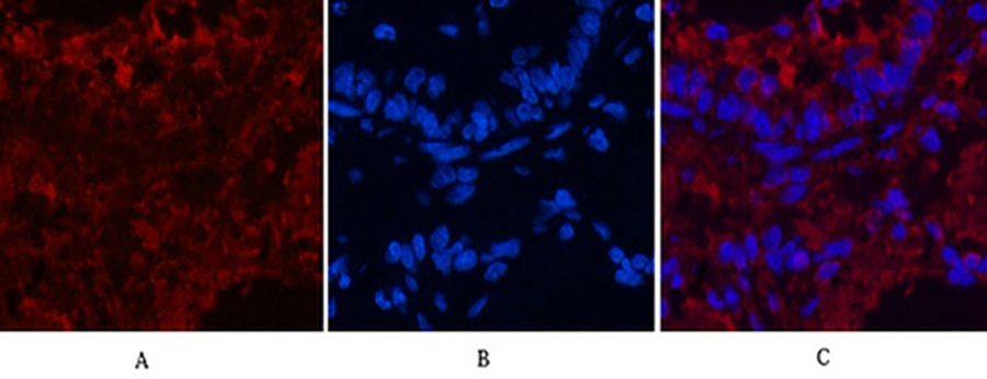
Fig.5. Immunofluorescence analysis of rat lung tissue. 1, CD4 Monoclonal Antibody (red) was diluted at 1:200 (4°C, overnight). 2, Cy3 Labeled secondary antibody was diluted at 1:300 (room temperature, 50min). 3, Picture B: DAPI (blue) 10min. Picture A: Target. Picture B: DAPI. Picture C: merge of A+B.
Author:Z Zhao, Q Zhao, B Gu Publication name:Theranostics IF:8
Author:LIU Yang, D Yahui, S Wei, DU Jiahui Publication name:Chinese Journal of Natural Medicines IF:5
Author:JH Kim, HJ Kim, DW Yoo, KD Park Publication name:Photodermatology, Photoimmunology & Photomedicine IF:3
You must be logged in to post a review.


Reviews
There are no reviews yet.