| Product name | VE-Cadherin Polyclonal Antibody |
| Immunogen | Synthesized peptide derived from the Internal region of human VE-Cadherin |
| Host | Rabbit |
| Reactivity | Human,Mouse,Rat |
| Applications | IF,WB,IHC,ELISA |
| Applications notes | Optimal working dilutions should be determined experimentally by the investigator. Suggested starting dilutions are as follows: IF 1:50-200;WB 1:500-2000;ELISA 1:10000-20000;IHC 1:50-300 |
| Clonality | Polyclonal |
| Preparation method | The antibody was affinity-purified from rabbit antiserum by affinity-chromatography using epitope-specific immunogen. |
| Alternative | CDH5; Cadherin-5; 7B4 antigen; Vascular endothelial cadherin; VE-cadherin; CD144 |
| Formulation | Liquid solution |
| Concentration | 1 mg/ml |
| Molecular weight | 110-130kD |
| Storage buffer | Liquid in PBS containing 50% glycerol, 0.5% BSA and 0.02% sodium azide. |
| Storage instructions | Stable for one year at -20°C from date of shipment. For maximum recovery of product, centrifuge the original vial after thawing and prior to removing the cap. Aliquot to avoid repeated freezing and thawing. |
| Shipping | Gel pack with blue ice. |
| Precautions | The product listed herein is for research use only and is not intended for use in human or clinical diagnosis. Suggested applications of our products are not recommendations to use our products in violation of any patent or as a license. We cannot be responsible for patent infringements or other violations that may occur with the use of this product. |
| Background | CDH5 encodes a classical cadherin of the cadherin superfamily. The encoded preproprotein is proteolytically processed to generate the mature glycoprotein. This calcium-dependent cell-cell adhesion molecule is comprised of five extracellular cadherin repeats, a transmembrane region and a highly conserved cytoplasmic tail. Functioning as a classical cadherin by imparting to cells the ability to adhere in a homophilic manner, this protein plays a role in endothelial adherens junction assembly and maintenance. CDH5 is located in a gene cluster in a region on the long arm of chromosome 16 that is involved in loss of heterozygosity events in breast and prostate cancer. |
| Gene ID | 1003 |
| Alternative | CDH5; Cadherin-5; 7B4 antigen; Vascular endothelial cadherin; VE-cadherin; CD144 |
| Others | VE-Cadherin Polyclonal Antibody detects endogenous levels of VE-Cadherin protein. |
| Accession | P33151 |
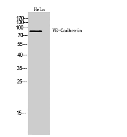
Fig.1. Western Blot analysis of Hela cells using VE-Cadherin Polyclonal Antibody. Antibody was diluted at 1:500.
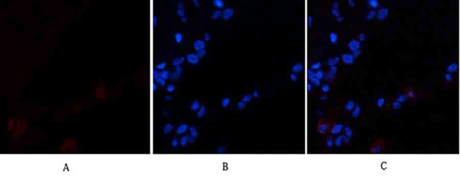
Fig.2. Immunofluorescence analysis of human lung tissue. 1, VE-Cadherin Polyclonal Antibody (red) was diluted at 1:200 (4°C, overnight). 2, Cy3 Labeled secondary antibody was diluted at 1:300 (room temperature, 50min). 3, Picture B: DAPI (blue) 10min. Picture A: Target. Picture B: DAPI. Picture C: merge of A+B.
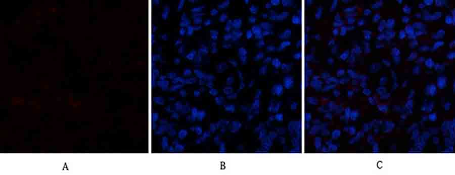
Fig.3. Immunofluorescence analysis of rat lung tissue. 1, VE-Cadherin Polyclonal Antibody (red) was diluted at 1:200 (4°C, overnight). 2, Cy3 Labeled secondary antibody was diluted at 1:300 (room temperature, 50min). 3, Picture B: DAPI (blue) 10min. Picture A: Target. Picture B: DAPI. Picture C: merge of A+B.
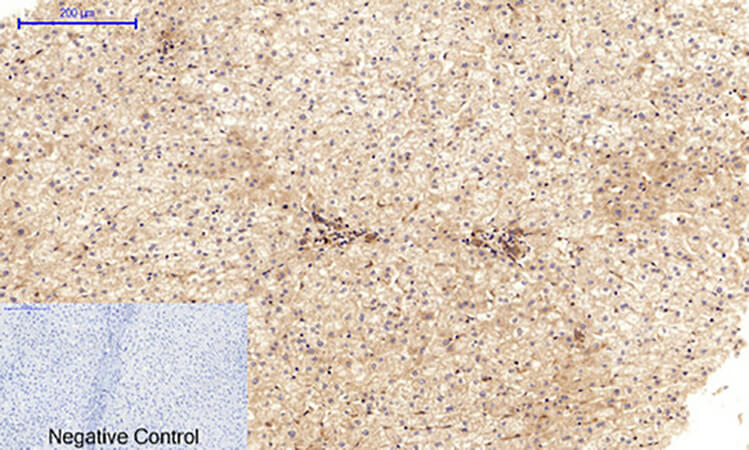
Fig.4. Immunohistochemical analysis of paraffin-embedded human liver tissue. 1, VE-Cadherin Polyclonal Antibody was diluted at 1:200 (4°C, overnight). 2, Sodium citrate pH 6.0 was used for antibody retrieval (>98°C, 20min). 3, secondary antibody was diluted at 1:200 (room temperature, 30min). Negative control was used by secondary antibody only.
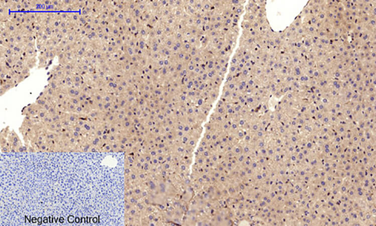
Fig.5. Immunohistochemical analysis of paraffin-embedded mouse liver tissue. 1, VE-Cadherin Polyclonal Antibody was diluted at 1:200 (4°C, overnight). 2, Sodium citrate pH 6.0 was used for antibody retrieval (>98°C, 20min). 3, secondary antibody was diluted at 1:200 (room temperature, 30min). Negative control was used by secondary antibody only.
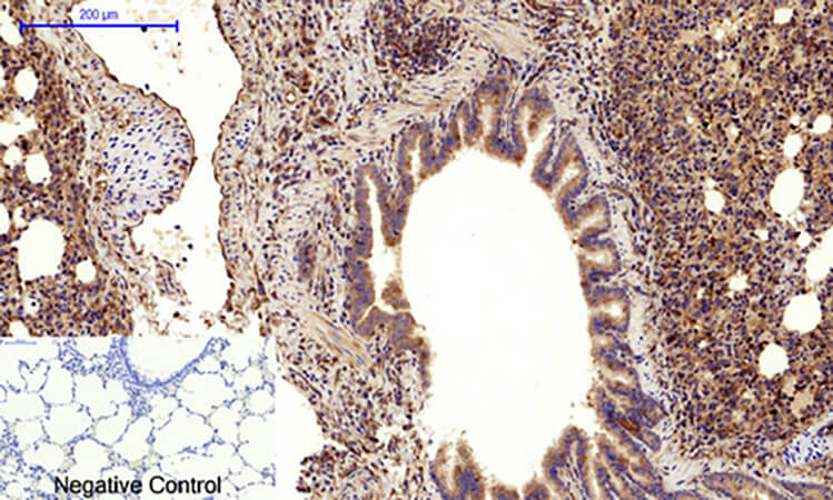
Fig.6. Immunohistochemical analysis of paraffin-embedded rat lung tissue. 1, VE-Cadherin Polyclonal Antibody was diluted at 1:200 (4°C, overnight). 2, Sodium citrate pH 6.0 was used for antibody retrieval (>98°C, 20min). 3, secondary antibody was diluted at 1:200 (room temperature, 30min). Negative control was used by secondary antibody only.
You must be logged in to post a review.


Reviews
There are no reviews yet.