| Product name | TraKine™ Pro Live-cell Tubulin-traker kit (Green Fluorescence) |
| Applications notes | TraKine™ Pro is series of long-term super-resolution cell staining imaging portfolio for labeling subcellular structures of live and fixed cells. |
| Kit components | •Tubulin Green (200 μM) •Buffer A (200 μM) •Buffer B (1 mM) |
| Features & Benefits | • Optimized staining protocol for labeling Tubulin in mammalian living cells. The product has been tested in U-2 OS,Hela,MCF-7 and MDA-MB-231 cell lines. U2OS cell line is preferred. If the sample type is not included in the above cell lines, we can provide experimental services for specific cell lines. • Suitable for Confocal high-definition imaging. • Suitable for structured light micro imaging (SIM) live cell research, ultra-high resolution microscopic imaging of live cells, and dynamic observation of cells in three-dimensional space. • Proprietary TubGreen™ (Ex/Em = 500/520 nm)-high specificity, low background and excellent photostability. • Low levels of cytotoxicity. |
| Usage notes | Make sure the pipette tips and PCR tubes were sterilized at high temperature and pressure. Make sure sterile environment and protect from light during the whole experiment. |
| Storage instructions | Refer to list of materials supplied for storage conditions of individual components. Stable for at least 6 months at recommended temperature from date of shipment. |
| Shipping | Gel pack with blue ice. |
| Precautions | The product listed herein is for research use only and is not intended for use in human or clinical diagnosis. Suggested applications of our products are not recommendations to use our products in violation of any patent or as a license. We cannot be responsible for patent infringements or other violations that may occur with the use of this product. |
| Background | Tubulin is the major building block of microtubules. This intracellular cylindrical filamentous structure is present in almost all eukaryotic cells. Microtubules function as structural and mobile elements in mitosis, intracellular transport, flagellar movement, and in the cytoskeleton. Tubulin is a heterodimer, which consists of a-tubulin and b-tubulin; both subunits have a molecular weight of 55 kDa and share considerable homology. The most studied tubulins have been isolated from vertebrate brains. The microtubules can be viewed in immunofluorescent microscopy, which enables the observation of the intracellular organization of proteins that are in the form of a supramolecular structure. |
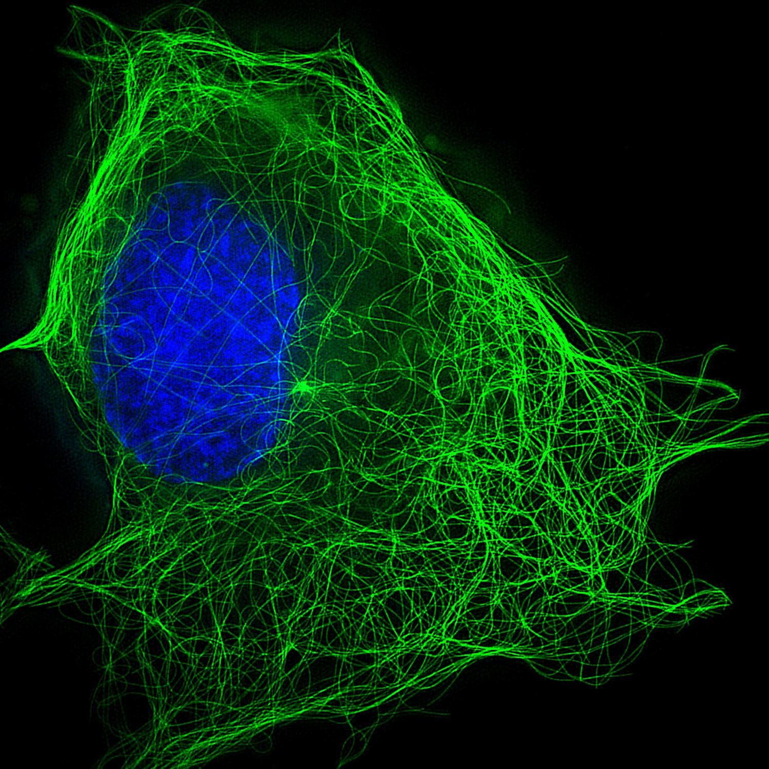
Fig.1. Markers of Tubulin -green in U-2 OS.
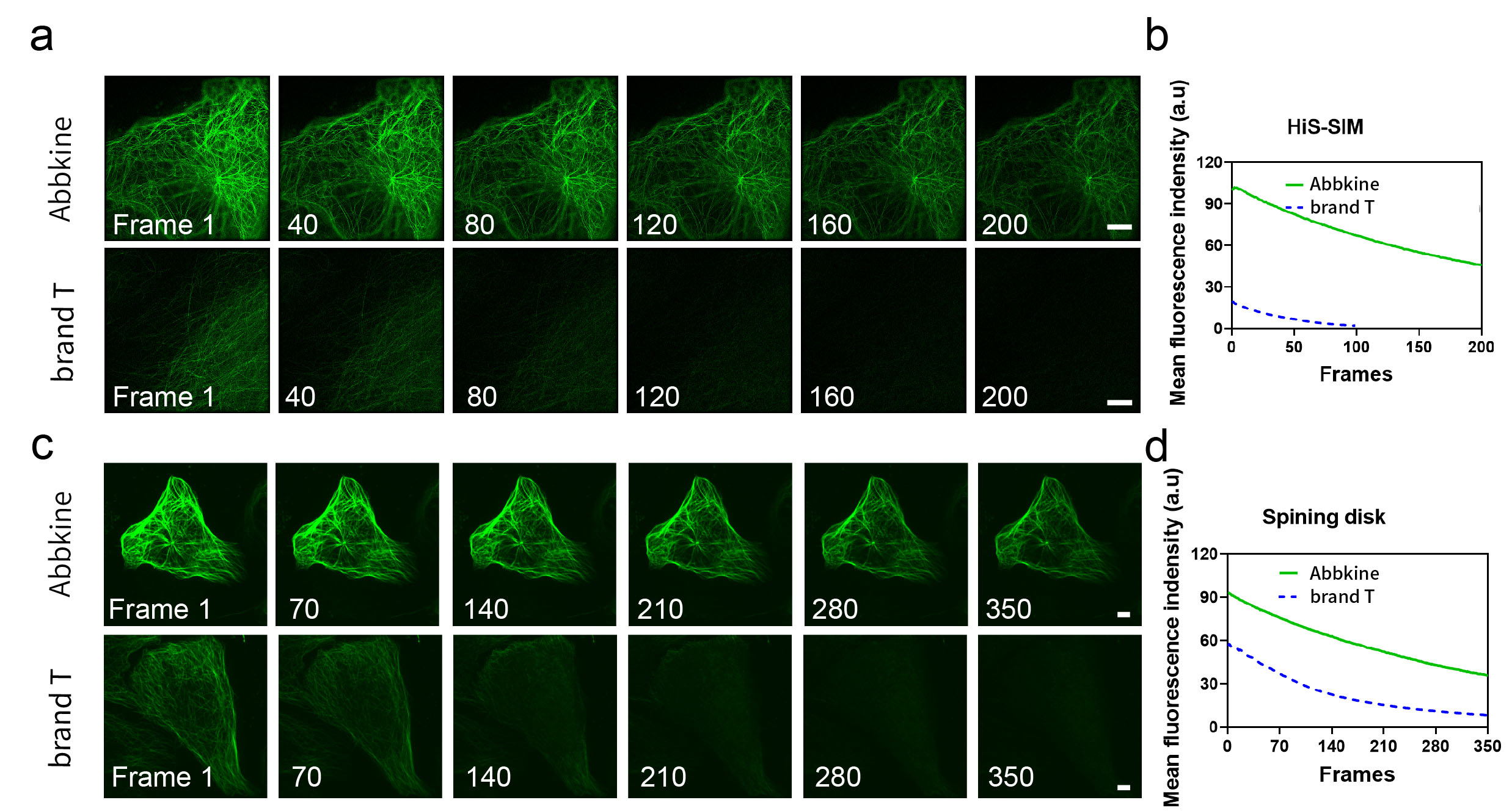
Fig.2. Long time series images (a, c) and corresponding fluorescence bleaching curves (b, d) were obtained by using HiS SIM Abbkine TraKine™ Pro Live-cell Tubulin-traker kit (Green Fluorescence) and Spining disk confocal microscopy of Tubulin Green and commercially available Tubulin Tracker Green on U-2 OS cells, respectively. Scale bars: 5 μM.
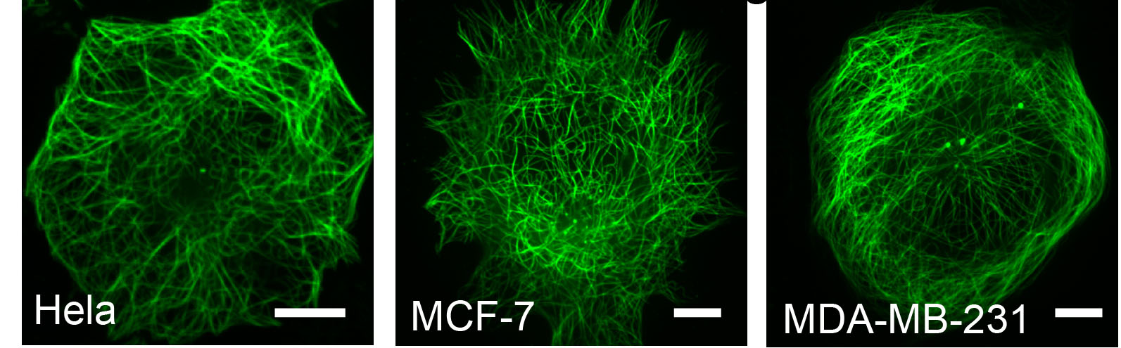
Fig.3. Confocal imaging results of other cells using Abbkine TraKine™ Pro Live-cell Tubulin-traker kit (Green Fluorescence).
You must be logged in to post a review.
1.The species of antibody reactivity should be the sample species that can be matched normally after Abbkine R&D experts have passed strict scientific verification. If your sample is not within the range of reactivity, in order to improve the efficiency and results of your experiment, it is not suggested to try other species. Otherwise, it may lead to sample mismatch and affect the effect of your experiment.
2.Please aliquot the antibody received as soon as possible and store it at -20℃, avoid repeated freezing and thawing, and use it within one year.
Welcome any form of communications, and better service will be provided here.
Tell: +1-404-854-0155
Email: service@abbkine.com
Support Email: support@abbkine.com
Address: 3052 Stroop Hill Road, Apt 203, Atlanta 30303, Georgia, United States of America


Reviews
There are no reviews yet.