| Product name | TGFβ1 Polyclonal Antibody |
| Immunogen | Synthesized peptide derived from the C-terminal region of human TGFβ1 at AA range: 310-390 |
| Host | Rabbit |
| Reactivity | Human, Mouse, Rat |
| Applications | ELISA, IF, IHC-P, WB |
| Applications notes | Optimal working dilutions should be determined experimentally by the investigator. Suggested starting dilutions are as follows: WB (1:500-1:2000), IHC-P (1:100-1:300), IF (1:200-1:1000), ELISA (1:20000). Not yet tested in other applications. |
| Clonality | Polyclonal |
| Preparation method | The antibody was affinity-purified from rabbit antiserum by affinity-chromatography using epitope-specific immunogen |
| Alternative | TGFB1; TGFB; Transforming growth factor beta-1; TGF-beta-1 |
| Formulation | Liquid solution |
| Concentration | 1 mg/ml |
| Storage buffer | PBS containing 50% Glycerol, 0.5% BSA and 0.02% Sodium Azide. |
| Storage instructions | Stable for one year at -20°C from date of shipment. For maximum recovery of product, centrifuge the original vial after thawing and prior to removing the cap. Aliquot to avoid repeated freezing and thawing. |
| Shipping | Gel pack with blue ice. |
| Precautions | The product listed herein is for research use only and is not intended for use in human or clinical diagnosis. Suggested applications of our products are not recommendations to use our products in violation of any patent or as a license. We cannot be responsible for patent infringements or other violations that may occur with the use of this product. |
| Background | TGFB1 encodes a secreted ligand of the TGF-beta (transforming growth factor-beta) superfamily of proteins. Ligands of this family bind various TGF-beta receptors leading to recruitment and activation of SMAD family transcription factors that regulate gene expression. The encoded preproprotein is proteolytically processed to generate a latency-associated peptide (LAP) and a mature peptide, and is found in either a latent form composed of a mature peptide homodimer, a LAP homodimer, and a latent TGF-beta binding protein, or in an active form consisting solely of the mature peptide homodimer. The mature peptide may also form heterodimers with other TGFB family members. The Transforming growth factor beta-1 regulates cell proliferation, differentiation and growth, and can modulate expression and activation of other growth factors including interferon gamma and tumor necrosis factor alpha. This gene is frequently upregulated in tumor cells, and mutations in this gene result in Camurati-Engelmann disease. |
| Gene ID | 7040 |
| Alternative | TGFB1; TGFB; Transforming growth factor beta-1; TGF-beta-1 |
| Others | TGFβ1 Polyclonal Antibody detects endogenous levels of TGFβ1 protein. |
| Accession | P01137 |
| Observed Band(KD) | 44 |
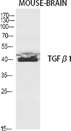
Fig.1. Western Blot analysis of various cells using TGFβ1 Polyclonal Antibody diluted at 1:2000.
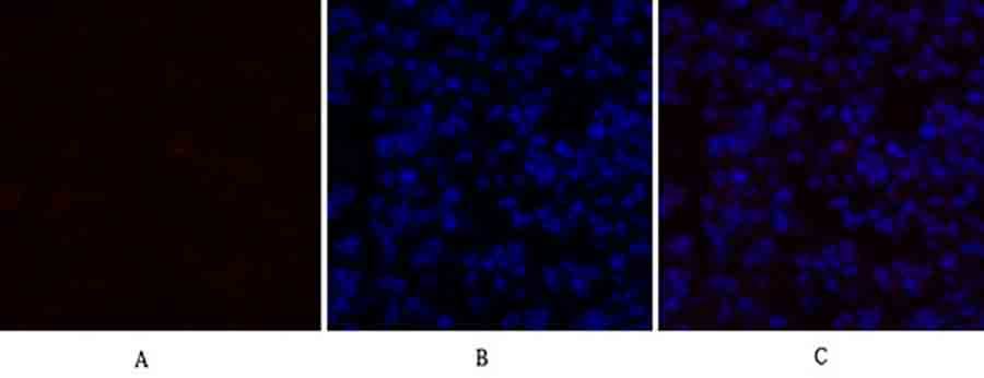
Fig.2. Immunofluorescence analysis of mouse lung tissue. 1, TGFβ1 Polyclonal Antibody (red) was diluted at 1:200 (4°C, overnight). 2, Cy3 Labeled secondary antibody was diluted at 1:300 (room temperature, 50min). 3, Picture B: DAPI (blue) 10min. Picture A: Target. Picture B: DAPI. Picture C: merge of A+B.
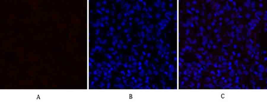
Fig.3. Immunofluorescence analysis of rat lung tissue. 1, TGFβ1 Polyclonal Antibody (red) was diluted at 1:200 (4°C, overnight). 2, Cy3 Labeled secondary antibody was diluted at 1:300 (room temperature, 50min). 3, Picture B: DAPI (blue) 10min. Picture A: Target. Picture B: DAPI. Picture C: merge of A+B.
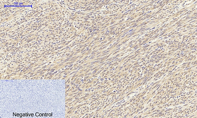
Fig.4. Immunohistochemical analysis of paraffin-embedded human uterus tissue. 1, TGFβ1 Polyclonal Antibody was diluted at 1:200 (4°C, overnight). 2, Sodium citrate pH 6.0 was used for antibody retrieval (>98°C, 20min). 3, secondary antibody was diluted at 1:200 (room temperature, 30min). Negative control was used by secondary antibody only.
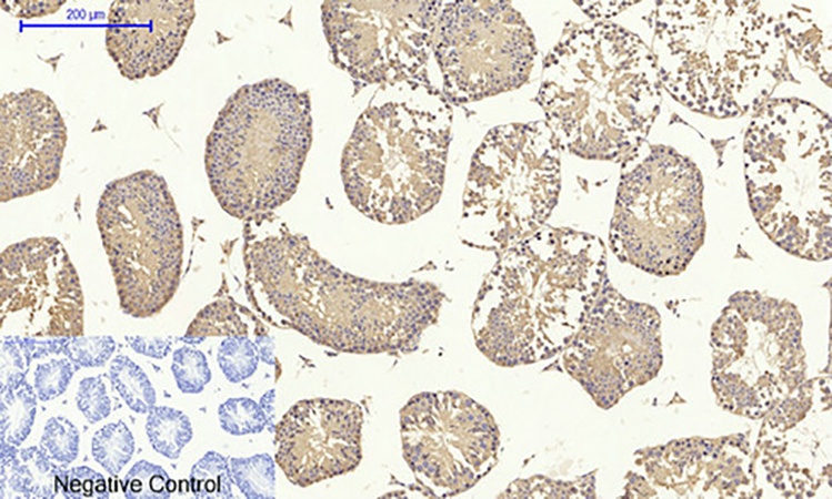
Fig.5. Immunohistochemical analysis of paraffin-embedded mouse testis tissue. 1, TGFβ1 Polyclonal Antibody was diluted at 1:200 (4°C, overnight). 2, Sodium citrate pH 6.0 was used for antibody retrieval (>98°C, 20min). 3, secondary antibody was diluted at 1:200 (room temperature, 30min). Negative control was used by secondary antibody only.
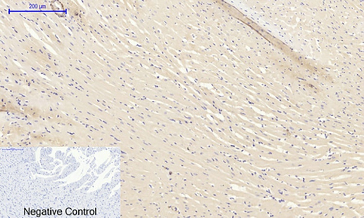
Fig.6. Immunohistochemical analysis of paraffin-embedded rat heart tissue. 1, TGFβ1 Polyclonal Antibody was diluted at 1:200 (4°C, overnight). 2, Sodium citrate pH 6.0 was used for antibody retrieval (>98°C, 20min). 3, secondary antibody was diluted at 1:200 (room temperature, 30min). Negative control was used by secondary antibody only.
Author:Zeng, Weiquan, et al. Publication name:Biomedicine & Pharmacotherapy IF:7.5
Author:Wen, Ying, et al. Publication name:Frontiers in Pharmacology IF:5.6
Author:Jia, Peizhi, et al. Publication name:Journal of Ethnopharmacology IF:5.4
You must be logged in to post a review.
1.The species of antibody reactivity should be the sample species that can be matched normally after Abbkine R&D experts have passed strict scientific verification. If your sample is not within the range of reactivity, in order to improve the efficiency and results of your experiment, it is not suggested to try other species. Otherwise, it may lead to sample mismatch and affect the effect of your experiment.
2.Please aliquot the antibody received as soon as possible and store it at -20℃, avoid repeated freezing and thawing, and use it within one year.
Welcome any form of communications, and better service will be provided here.
Tell: +1-404-854-0155
Email: service@abbkine.com
Support Email: support@abbkine.com
Address: 3052 Stroop Hill Road, Apt 203, Atlanta 30303, Georgia, United States of America


Reviews
There are no reviews yet.